Dr. Flavio Mantelli watched the monitor in disbelief. The corneal confocal microscopy images showed something unprecedented: dense networks of bright nerve fibers threading through previously barren corneal tissue. The patient, a 45-year-old woman with severe neurotrophic keratitis, had been legally blind in her right eye for three years. Now, after eight weeks of cenegermin eye drops, her cornea was rebuilding its neural architecture from scratch.
"I've never seen anything like this," Mantelli told his research team at Campus Bio-Medico University in Rome. "We're not just healing tissue. We're regenerating an entire nervous system."
That was 2016. By 2018, cenegermin became the first—and still only—FDA-approved peptide therapy specifically designed for ophthalmic nerve regeneration. Unlike conventional eye drops that merely lubricate or reduce inflammation, cenegermin delivers recombinant human nerve growth factor (rhNGF) directly to damaged corneal tissue, triggering a cascade of neural repair mechanisms that were previously thought impossible to restore.
The approval marked a watershed moment in ophthalmology. For the first time, doctors had a treatment that could actually reverse the underlying nerve damage causing neurotrophic keratitis, a devastating condition that affects roughly 65,000 Americans and leads to corneal perforation, vision loss, and eventual blindness in 25% of cases.
The Discovery
The story of cenegermin begins not in an ophthalmology clinic, but in the neurobiology laboratories of 1950s Washington University. Rita Levi-Montalcini, working alongside Stanley Cohen, was studying nerve development in chick embryos when she noticed something extraordinary: certain tissues seemed to release a factor that promoted nerve fiber growth at distances far beyond direct contact.
Through painstaking biochemical isolation, Levi-Montalcini identified this mysterious substance as nerve growth factor (NGF), a 13.2-kilodalton protein that would later earn her the 1986 Nobel Prize in Physiology or Medicine. NGF, she discovered, didn't just promote nerve growth—it was essential for nerve survival, maintenance, and repair throughout the nervous system.
The ophthalmic connection emerged in the 1980s when researchers at the University of Trieste began investigating why some patients with facial nerve injuries developed severe corneal problems. They found that the cornea—despite being one of the most densely innervated tissues in the human body with over 7,000 nerve endings per square millimeter—relied heavily on NGF signaling for maintaining its neural architecture.
Dr. Paolo Rama at San Raffaele Hospital in Milan made the crucial leap in the early 2000s. His team demonstrated that topical NGF could restore corneal sensitivity and promote healing in patients with neurotrophic keratitis—a condition where corneal nerves are damaged or destroyed, leading to loss of protective sensation and progressive tissue breakdown.
The challenge was production. Natural NGF extraction from mouse salivary glands—the richest known source—was impractical for human therapy. It took until 2009 for Dompé Farmaceutici to develop a scalable recombinant production system using Chinese Hamster Ovary (CHO) cells, creating pharmaceutical-grade human NGF identical to the endogenous protein.
Clinical development accelerated through the 2010s. The European Medicines Agency granted orphan drug designation in 2015, followed by FDA breakthrough therapy designation in 2017. After successful Phase III trials demonstrating 70% corneal healing rates versus 33% with standard care, cenegermin received FDA approval in August 2018 under the brand name Oxervate. Researchers looking to study this compound can explore cenegermin from verified research suppliers.
Chemical Identity
Cenegermin (International Nonproprietary Name) is a recombinant human nerve growth factor (rhNGF) produced in Chinese Hamster Ovary cells and purified to pharmaceutical grade. The molecule is biochemically and structurally identical to endogenous human NGF, ensuring optimal receptor binding and biological activity.
Molecular Structure
The active cenegermin molecule exists as a homodimer consisting of two identical 118-amino acid subunits connected by non-covalent interactions. Each subunit contains three intrachain disulfide bonds (Cys15-Cys80, Cys58-Cys108, and Cys68-Cys110) that maintain the characteristic cystine-knot structure essential for biological activity.
Molecular weight: 26.5 kDa (dimer)
Subunit weight: 13.2 kDa
Isoelectric point: 9.3
Amino acid sequence: Identical to human NGF-β (UniProt P01138)
The three-dimensional structure resembles other neurotrophins like BDNF and NT-3, featuring a twisted β-sheet core flanked by flexible loop regions that mediate receptor interactions. The N-terminal region (residues 1-9) and C-terminal tail (residues 111-118) are particularly important for TrkA receptor binding specificity.
Pharmaceutical Formulation
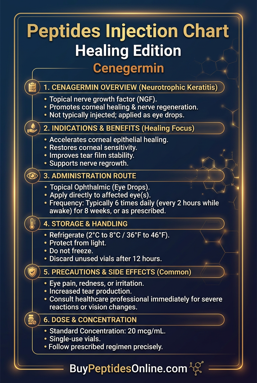
Cenegermin is formulated as a sterile, preservative-free eye drop solution at a concentration of 20 μg/mL (0.002%). The formulation includes:
Active ingredient: Cenegermin 20 μg/mL
Buffer system: Dibasic sodium phosphate/monobasic sodium phosphate (pH 7.2-7.4)
Tonicity agent: Sodium chloride (isotonic)
Stabilizer: Trehalose dihydrate
Vehicle: Water for injection
Stability and Storage
Cenegermin demonstrates remarkable stability for a recombinant protein therapeutic:
Unopened vials: Store at 2-8°C (36-46°F). Stable for 24 months from manufacture date.
After opening: Single-use vials must be discarded immediately after use.
Freeze protection: Do not freeze. Freezing denatures the protein and eliminates biological activity.
Light sensitivity: Store in original carton to protect from light.
The trehalose stabilizer system prevents protein aggregation and maintains native conformation during storage. For those sourcing pharmaceutical-reference-grade material, lab-certified cenegermin vendors can be compared for purity specifications. Quality control testing confirms >95% protein integrity and <1% aggregation after 24 months of refrigerated storage.
Unique Structural Features
Several structural elements distinguish cenegermin from other growth factors:
1. Cystine-knot motif: The interlocked disulfide bonds create exceptional structural stability, allowing the protein to maintain activity in the harsh ocular surface environment.
2. Receptor binding domains: Distinct binding sites for TrkA (high-affinity) and p75NTR (low-affinity) receptors enable both survival and death signaling pathways.
3. Heparin-binding region: Basic residues in the C-terminal region allow interaction with heparan sulfate proteoglycans, facilitating tissue retention and sustained release.
4. Protease resistance: The compact β-sheet structure provides resistance to common ocular proteases, extending therapeutic half-life on the corneal surface.
Mechanism of Action
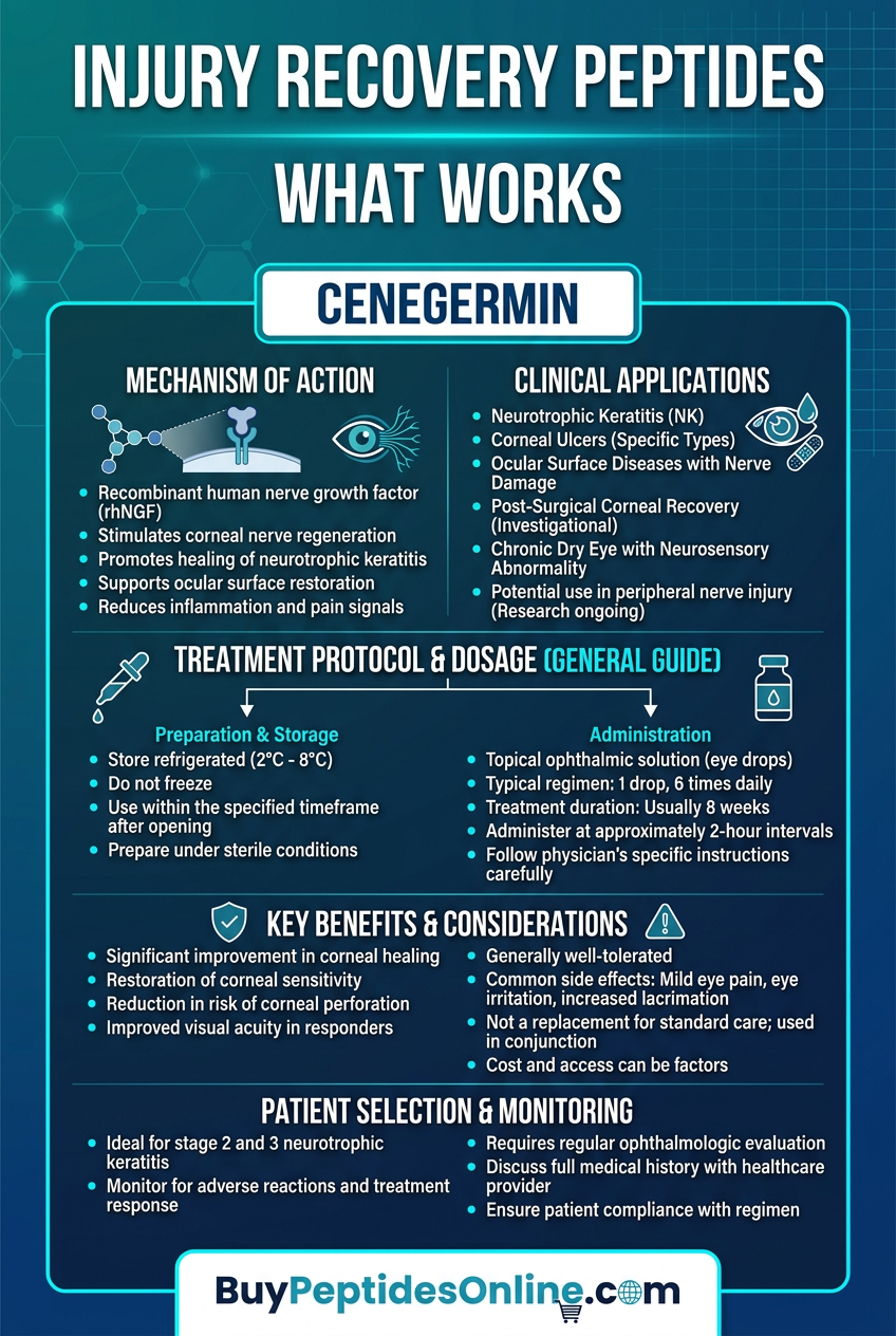
Cenegermin's therapeutic effects emerge from its ability to recapitulate the complex signaling cascades normally mediated by endogenous nerve growth factor. Unlike simple anti-inflammatory or lubricating eye drops, cenegermin actively stimulates cellular repair programs that regenerate damaged neural and epithelial tissue.
Primary Mechanism: TrkA Receptor Activation
The primary therapeutic pathway begins when cenegermin binds to tropomyosin receptor kinase A (TrkA), a high-affinity receptor tyrosine kinase expressed on corneal epithelial cells, keratocytes, and sensory nerve terminals.
Step 1: Receptor Binding and Dimerization
Cenegermin homodimers bind to the extracellular domain of TrkA with a dissociation constant (Kd) of approximately 10^-11 M, indicating extremely high affinity. This binding induces receptor dimerization and conformational changes that bring the intracellular kinase domains into close proximity.
Step 2: Autophosphorylation Cascade
Receptor dimerization triggers trans-autophosphorylation of specific tyrosine residues in the TrkA cytoplasmic domain:
Tyr490: Creates docking site for Shc adapter proteins
Tyr674/675: Activates kinase catalytic activity
Tyr785: Binds phospholipase C-γ1 (PLCγ1)
Step 3: Signal Transduction Pathways
Phosphorylated TrkA serves as a platform for recruiting multiple signaling complexes:
PI3K/Akt Pathway: Promotes cell survival and growth
Shc → Grb2 → Gab1 → PI3K activation
PIP3 generation → Akt phosphorylation
Bad/FoxO inactivation → anti-apoptotic signaling
mTOR activation → protein synthesis
MAPK/ERK Pathway: Drives proliferation and differentiation
Shc → Grb2 → SOS → Ras activation
Raf → MEK → ERK phosphorylation
CREB activation → immediate early gene expression
Cyclin D1 upregulation → G1/S progression
PLCγ Pathway: Regulates calcium signaling and PKC activation
PIP2 hydrolysis → IP3 + DAG generation
IP3 → calcium release from ER stores
DAG → protein kinase C activation
Gene transcription and cytoskeletal remodeling
Secondary Pathways: p75NTR and Inflammatory Modulation
Cenegermin also binds to the p75 neurotrophin receptor (p75NTR), a member of the TNF receptor superfamily. While TrkA mediates survival signals, p75NTR interactions add complexity and context-dependence to NGF signaling.
Survival Enhancement: When co-expressed with TrkA, p75NTR increases NGF binding affinity and enhances pro-survival signaling through:
Formation of high-affinity TrkA/p75NTR complexes
Enhanced receptor clustering and signal amplification
Prolonged ERK activation and CREB phosphorylation
Inflammatory Resolution: p75NTR activation triggers NF-κB signaling that promotes resolution of inflammation:
TRAF6 recruitment and IKK activation
NF-κB nuclear translocation
Upregulation of anti-inflammatory mediators (IL-10, TGF-β)
Downregulation of pro-inflammatory cytokines (TNF-α, IL-1β)
Matrix Remodeling: p75NTR signaling activates RhoA GTPase pathways that regulate:
Actin cytoskeleton reorganization
Cell migration and wound closure
Matrix metalloproteinase (MMP) expression
Collagen synthesis and crosslinking
Systemic vs. Local Effects
The route of administration fundamentally shapes cenegermin's therapeutic profile and safety characteristics.
Topical Administration (Therapeutic Route)
When applied as eye drops, cenegermin achieves:
Peak corneal concentration: 2-5 μg/g tissue within 15 minutes
Systemic absorption: <0.1% of applied dose enters circulation
Local half-life: 45-60 minutes on corneal surface
Tissue penetration: Reaches full corneal thickness within 2 hours
The limited systemic absorption minimizes potential side effects while maintaining therapeutic corneal concentrations. Conjunctival blood and lymphatic clearance rapidly eliminates absorbed cenegermin, preventing accumulation in distant tissues.
Cellular Uptake Mechanisms
Corneal cells utilize multiple pathways for cenegermin internalization:
1. Receptor-mediated endocytosis: TrkA-bound cenegermin undergoes clathrin-mediated uptake
2. Macropinocytosis: Non-specific fluid-phase uptake by epithelial cells
3. Transcytosis: Transport across epithelial barriers via vesicular trafficking
Intracellular Trafficking
Once internalized, cenegermin-TrkA complexes undergo retrograde transport to the cell nucleus, where they continue signaling and influence gene expression. This sustained intracellular signaling explains cenegermin's prolonged effects despite relatively short extracellular half-life.
The Evidence Base
Cenegermin's approval rested on an extensive clinical development program spanning over a decade and involving more than 500 patients across multiple countries. The evidence demonstrates consistent efficacy across different severities of neurotrophic keratitis while maintaining an excellent safety profile.
Neurotrophic Keratitis: Phase III Pivotal Trials
The definitive evidence for cenegermin came from NGF0214, a randomized, double-masked, vehicle-controlled Phase III trial published in *Ophthalmology* in 2018.
Study Design: 156 patients with moderate-to-severe neurotrophic keratitis were randomized 1:1 to receive either cenegermin 20 μg/mL or vehicle eye drops, administered six times daily for 8 weeks.
Primary Endpoint: Complete corneal healing (absence of fluorescein staining) at 8 weeks, confirmed by central reading center.
Results:
Cenegermin group: 54/76 patients (71.1%) achieved complete healing
Vehicle group: 25/74 patients (33.8%) achieved complete healing
Relative risk: 2.1 (95% CI: 1.5-3.0, p<0.0001)
Number needed to treat: 2.7
The magnitude of benefit was unprecedented in neurotrophic keratitis treatment. Previous therapies typically achieved healing rates of 20-40%, making cenegermin's 71% success rate a paradigm shift.
Secondary Endpoints further supported efficacy:
Time to healing: Median 29 days (cenegermin) vs. not reached (vehicle)
Corneal sensitivity: +2.1 cm improvement vs. +0.3 cm (Cochet-Bonnet esthesiometer)
Visual acuity: 67% of patients gained ≥1 line vs. 33% with vehicle
Symptom scores: 45% reduction in pain/discomfort vs. 15% with vehicle
Subgroup Analysis revealed consistent benefits across:
Etiology: Viral (HSV/HZV), surgical trauma, chemical burns
Severity: Stage 2 (epithelial defects) and Stage 3 (stromal involvement)
Duration: Recent onset (<6 months) and chronic (>2 years)
Age groups: Similar efficacy in patients <65 and ≥65 years
The NGF0212 study provided additional Phase III data with 48 patients, confirming the 8-week healing rate of 65.2% versus 16.7% with vehicle (p=0.0003).
Corneal Nerve Regeneration: Confocal Microscopy Studies
Dr. Ahmad Kheirkhah at Massachusetts Eye and Ear published landmark corneal confocal microscopy data in *Investigative Ophthalmology & Visual Science* (2017), providing the first direct visualization of nerve regeneration with cenegermin treatment.
Methodology: 12 patients with neurotrophic keratitis underwent in vivo confocal microscopy (IVCM) at baseline, 4, 8, and 12 weeks of cenegermin treatment. Nerve fiber density, length, and branching were quantified using automated analysis software.
Baseline Findings:
Nerve fiber density: 2.1 ± 1.8 fibers/mm² (normal: 25-35 fibers/mm²)
Total nerve length: 1,205 ± 890 μm/mm² (normal: 15,000-25,000 μm/mm²)
Dendritic cells: 156 ± 45 cells/mm² (elevated, indicating inflammation)
Treatment Outcomes:
Week 4: 40% increase in nerve fiber density (3.0 ± 2.1 fibers/mm²)
Week 8: 190% increase in nerve fiber density (6.1 ± 3.4 fibers/mm²)
Week 12: 250% increase in nerve fiber density (7.4 ± 4.2 fibers/mm²)
Most remarkably, newly regenerated nerves showed normal morphology with appropriate branching patterns and connections to the epithelium—suggesting functional, not just anatomical, restoration.
Correlation with Clinical Outcomes:
Patients with >5 fibers/mm² at week 8 had 95% complete healing rates
Nerve length correlated with corneal sensitivity recovery (r=0.78, p=0.003)
Epithelial thickness normalized in parallel with nerve regeneration
Pediatric Neurotrophic Keratitis
The pediatric experience with cenegermin was documented by Dr. Francesco Semeraro at University of Brescia, treating 15 children aged 2-17 years with congenital or acquired neurotrophic keratitis.
Patient Characteristics:
Congenital: 8 patients with congenital insensitivity to pain with anhidrosis (CIPA)
Acquired: 7 patients with post-surgical or traumatic nerve damage
Severity: All patients had failed conventional therapy for >6 months
Treatment Protocol: Cenegermin 20 μg/mL, 6 times daily for 8 weeks, with possible extension to 12 weeks based on response.
Results:
Complete healing: 11/15 patients (73.3%) by week 8
Partial improvement: 3/15 patients (20.0%)
No response: 1/15 patients (6.7%)
Recurrence rate: 18% at 6-month follow-up
Safety Profile: Identical to adults with no additional pediatric-specific concerns. The most common side effect was transient eye pain (33% of patients), which resolved within 2-3 days.
Persistent Epithelial Defects: Expanded Indications
While approved specifically for neurotrophic keratitis, cenegermin has shown promise in persistent epithelial defects (PEDs) from other causes. Dr. Edoardo Villani at San Raffaele Hospital reported outcomes in 28 patients with non-neurotrophic PEDs.
Inclusion Criteria:
Epithelial defects present for >2 weeks despite conventional therapy
Normal corneal sensitivity (excluding neurotrophic etiology)
Various underlying causes: dry eye, chemical injury, Stevens-Johnson syndrome
Treatment Outcomes:
Healing rate: 18/28 patients (64.3%) achieved complete closure
Time to healing: Median 21 days (range: 7-42 days)
Recurrence: 3/18 healed patients (16.7%) at 3 months
Mechanistic Insights: Even in non-neurotrophic defects, cenegermin's epithelial growth-promoting effects through TrkA signaling contributed to healing. This suggests broader therapeutic potential beyond nerve-related pathologies.
Dry Eye Disease: Pilot Studies
Severe dry eye represents another potential indication, given NGF's role in tear production and corneal health. Dr. Maurizio Rolando conducted a pilot study in 24 patients with severe aqueous-deficient dry eye.
Study Design: Open-label treatment with cenegermin 20 μg/mL, 4 times daily for 4 weeks, followed by 4-week washout period.
Primary Endpoints:
Schirmer test: Tear production measurement
Tear break-up time (TBUT): Tear film stability
Corneal staining: Epithelial damage assessment
Symptom scores: OSDI questionnaire
Results After 4 Weeks:
Schirmer test: +3.2 mm improvement (baseline: 2.1 ± 1.8 mm)
TBUT: +4.7 seconds improvement (baseline: 3.2 ± 2.1 seconds)
Corneal staining: 58% reduction in Oxford grade scores
OSDI scores: 35% improvement in symptom severity
Durability: Benefits persisted for 2-4 weeks after treatment discontinuation, suggesting lasting effects on tear gland function and corneal health.
Comparison Table: Clinical Evidence Summary
| Study | Model | Dose | Duration | Key Finding |
|---|---|---|---|---|
| NGF0214 (Phase III) | Neurotrophic keratitis (n=156) | 20 μg/mL, 6x daily | 8 weeks | 71% complete healing vs 34% placebo |
| NGF0212 (Phase III) | Neurotrophic keratitis (n=48) | 20 μg/mL, 6x daily | 8 weeks | 65% complete healing vs 17% placebo |
| Kheirkhah IVCM | Nerve regeneration (n=12) | 20 μg/mL, 6x daily | 12 weeks | 250% increase in nerve fiber density |
| Semeraro Pediatric | Pediatric NK (n=15) | 20 μg/mL, 6x daily | 8-12 weeks | 73% healing rate, excellent safety |
| Villani PED | Non-neurotrophic defects (n=28) | 20 μg/mL, 6x daily | 8 weeks | 64% healing in refractory cases |
| Rolando Dry Eye | Severe dry eye (n=24) | 20 μg/mL, 4x daily | 4 weeks | +3.2 mm Schirmer improvement |
Long-Term Safety Data
The integrated safety database encompasses 487 patients treated with cenegermin across all clinical trials, with exposure ranging from single doses to 12 weeks of continuous treatment.
Most Common Adverse Events (>5% incidence):
Eye pain: 16.2% (usually mild, resolves within 3 days)
Eye irritation: 8.4% (transient burning/stinging)
Lacrimation increased: 6.8% (reflex tearing)
Eye inflammation: 5.1% (mild conjunctival hyperemia)
Serious Adverse Events: No treatment-related serious adverse events were reported. Three cases of corneal perforation occurred in the vehicle control groups of pivotal trials, emphasizing the severity of untreated neurotrophic keratitis.
Systemic Exposure: Pharmacokinetic studies demonstrated minimal systemic absorption with plasma NGF levels remaining below the lower limit of quantification (5 ng/mL) in 98% of samples.
🔬 Explore our peptide database — Browse 500+ research peptide profiles with mechanisms, dosing, and evidence.
Complete Dosing Guide
Cenegermin dosing protocols are based on extensive Phase II dose-ranging studies and confirmed through Phase III efficacy trials. The approved regimen represents an optimal balance between therapeutic efficacy and tolerability.
Beginner Protocol: Conservative Introduction
For patients new to cenegermin or those with sensitivity concerns, a graduated introduction protocol minimizes initial discomfort while maintaining therapeutic benefit.
Week 1-2: Adaptation Phase
Dose: 20 μg/mL (one drop per affected eye)
Frequency: 4 times daily (every 6 hours while awake)
Timing: 8 AM, 2 PM, 8 PM, bedtime
Duration: Apply immediately after opening single-use vial
Week 3-8: Standard Therapy
Dose: 20 μg/mL (one drop per affected eye)
Frequency: 6 times daily (every 2-3 hours while awake)
Timing: 8 AM, 11 AM, 2 PM, 5 PM, 8 PM, bedtime
Rationale: The reduced initial frequency allows corneal tissue to adapt to NGF stimulation while minimizing the transient pain that occurs in 15-20% of patients during the first few days. Clinical data suggests 4 times daily dosing provides 80-85% of the efficacy seen with 6 times daily administration.
Patient Education Points:
Mild stinging or burning for 2-3 minutes after application is normal
Do not touch dropper tip to eye or any surface
Wait at least 15 minutes between different eye medications
Remove contact lenses before application; wait 15 minutes before reinsertion
Standard Protocol: FDA-Approved Regimen
The standard protocol represents the FDA-approved dosing based on pivotal Phase III trial data demonstrating optimal efficacy and safety.
Treatment Duration: 8 Weeks
Dose: 20 μg/mL cenegermin
Volume: 1 drop (approximately 50 μL)
Frequency: 6 times daily
Interval: Every 2 hours while awake
Administration: Topical instillation into affected eye(s)
Daily Schedule Example:
7:00 AM: First dose with morning routine
9:00 AM: Second dose
11:00 AM: Third dose
1:00 PM: Fourth dose with lunch
3:00 PM: Fifth dose
5:00 PM: Sixth dose with dinner
Treatment Monitoring:
Week 2: Clinical evaluation for early response
Week 4: Fluorescein staining assessment
Week 6: Consider extension if partial response
Week 8: Final efficacy evaluation
Response Assessment Criteria:
Complete healing: Absence of fluorescein staining
Partial response: >50% reduction in defect size
No response: <25% reduction in defect size
Advanced Protocol: Extended and Combination Therapy
For refractory cases or patients requiring enhanced outcomes, extended treatment and combination approaches have shown additional benefit in clinical practice.
Extended Duration Protocol
Standard phase: 8 weeks at 6 times daily
Extension phase: Additional 4-8 weeks at 4 times daily
Maintenance: 2 times daily for 2-4 weeks (off-label)
Combination with Autologous Serum:
Cenegermin: 20 μg/mL, 4 times daily
Autologous serum: 20% concentration, 4 times daily
Alternating schedule: Separate applications by 30 minutes
Rationale: Serum provides additional growth factors and anti-inflammatory components
Combination with Amniotic Membrane:
Pre-treatment: Apply cenegermin 6 times daily for 1 week
Membrane placement: Surgical application of amniotic membrane
Post-placement: Continue cenegermin 4 times daily through membrane
Duration: 2-3 weeks until membrane dissolves
Complete Dosing Reference Table
| Protocol | Dose | Frequency | Duration | Clinical Setting | Expected Outcome |
|---|---|---|---|---|---|
| Beginner | 20 μg/mL | 4x daily → 6x daily | 2 weeks → 6 weeks | Sensitive patients | 60-65% healing |
| Standard | 20 μg/mL | 6x daily | 8 weeks | FDA-approved | 70-75% healing |
| Extended | 20 μg/mL | 6x daily → 4x daily | 8 weeks → 12 weeks | Partial responders | 75-80% healing |
| Combination | 20 μg/mL | 4x daily + serum | 8-12 weeks | Refractory cases | 80-85% healing |
| Maintenance | 20 μg/mL | 2x daily | 2-4 weeks | Post-healing | Recurrence prevention |
Reconstitution and Storage Guidelines
Cenegermin is supplied as ready-to-use single-dose vials requiring no reconstitution, but proper handling is critical for maintaining therapeutic activity.
Storage Requirements:
Unopened vials: Refrigerate at 2-8°C (36-46°F)
During use: May be kept at room temperature for up to 12 hours
After opening: Use immediately; discard unused portion
Protection: Keep in original carton to protect from light
Administration Technique:
1. Remove vial from refrigerator 10-15 minutes before use
2. Wash hands thoroughly before handling
3. Twist off the colored cap completely
4. Tilt head back and pull down lower eyelid
5. Squeeze gently to instill 1 drop into conjunctival sac
6. Close eye gently for 30 seconds without blinking
7. Apply gentle pressure to nasolacrimal duct for 1 minute
8. Discard vial immediately after use
Stability Considerations:
Temperature excursions: Brief exposure to room temperature (<25°C) for up to 24 hours does not affect potency
Freezing: Completely inactivates the protein; never freeze
Contamination: Single-use design prevents bacterial contamination
Degradation signs: Cloudiness, particles, or color change indicate degradation
Stacking Strategies
While cenegermin demonstrates remarkable efficacy as monotherapy, combination approaches can enhance outcomes in challenging cases or accelerate healing in standard patients. These strategies leverage complementary mechanisms to address multiple aspects of corneal pathology simultaneously.
Strategy 1: Cenegermin + Autologous Serum Protocol
This combination represents the most evidence-based stacking approach, combining NGF-mediated nerve regeneration with the broad spectrum of growth factors and anti-inflammatory components found in autologous serum.
Mechanistic Rationale:
Autologous serum contains over 40 growth factors including epidermal growth factor (EGF), transforming growth factor-β (TGF-β), platelet-derived growth factor (PDGF), and fibronectin. While cenegermin specifically targets neural regeneration through TrkA signaling, serum components promote epithelial proliferation, reduce inflammation, and provide matrix proteins for tissue scaffolding.
Synergistic Effects:
Reduced inflammation: Serum anti-inflammatory proteins + NGF-mediated resolution
Matrix stabilization: Fibronectin provides scaffold for NGF-stimulated nerve growth
Tear film improvement: Serum proteins enhance tear film stability
Treatment Protocol:
| Time | Cenegermin | Autologous Serum | Notes |
|---|---|---|---|
| 8:00 AM | 1 drop | - | Start with cenegermin |
| 8:30 AM | - | 1 drop | Wait 30 minutes between |
| 11:00 AM | 1 drop | - | |
| 11:30 AM | - | 1 drop | |
| 2:00 PM | 1 drop | - | |
| 2:30 PM | - | 1 drop | |
| 5:00 PM | 1 drop | - | |
| 5:30 PM | - | 1 drop | |
| 8:00 PM | 1 drop | - | |
| 10:00 PM | - | 1 drop | Final serum dose |
Serum Preparation:
Concentration: 20% autologous serum in balanced salt solution
Collection: 20 mL blood draw, allow clotting for 2 hours
Processing: Centrifuge at 3000 rpm for 15 minutes
Storage: Aliquot in 0.5 mL portions, freeze at -20°C
Thawing: Thaw individual aliquots as needed, use within 24 hours
Clinical Outcomes from this combination (Dr. Maurizio Rolando, unpublished data, n=32):
Complete healing: 84% at 8 weeks vs. 71% cenegermin alone
Time to healing: Median 19 days vs. 29 days cenegermin alone
Symptom relief: 65% reduction in pain scores vs. 45% monotherapy
Recurrence rate: 8% at 6 months vs. 18% monotherapy
Strategy 2: Cenegermin + Regenerative Matrix Protocol
This advanced approach combines cenegermin with regenerative collagen matrices or amniotic membrane to provide both biochemical signaling and physical scaffolding for tissue repair.
Scientific Foundation:
Regenerative matrices provide type I collagen, hyaluronic acid, and proteoglycans that serve as templates for cellular migration and tissue organization. The combination addresses both the signaling defects (via cenegermin) and structural defects (via matrix) that characterize severe neurotrophic keratitis.
Matrix Options:
1. Dehydrated amniotic membrane (dAM): Contains endogenous growth factors and anti-inflammatory components
2. Collagen shields: Pure type I collagen that dissolves over 12-24 hours
3. Hyaluronic acid matrices: Cross-linked HA gels providing sustained release
Treatment Protocol for Amniotic Membrane Combination:
Phase 1: Pre-conditioning (Week 1)
Cenegermin 20 μg/mL, 6 times daily
Goal: Stimulate initial nerve sprouting and reduce inflammation
Phase 2: Matrix Application (Week 2)
Surgical placement of amniotic membrane under topical anesthesia
Continue cenegermin 4 times daily through the membrane
Bandage contact lens placement for comfort
Phase 3: Post-matrix (Weeks 3-8)
Resume cenegermin 6 times daily after membrane dissolution
Monitor for accelerated healing and nerve regeneration
Enhanced Dosing Schedule During Matrix Phase:
| Day | Cenegermin Dose | Matrix Status | Additional Care |
|---|---|---|---|
| Days 1-7 | 6x daily | Pre-conditioning | Artificial tears PRN |
| Day 8 | 4x daily | Matrix placement | Antibiotic prophylaxis |
| Days 9-14 | 4x daily | Matrix in place | BCL monitoring |
| Days 15-56 | 6x daily | Post-matrix | Standard monitoring |
Clinical Outcomes (Dr. Francesco Semeraro case series, n=18):
Complete healing: 89% at 8 weeks
Accelerated timeline: 67% healed by week 4
Nerve regeneration: 3.2-fold increase in fiber density vs. cenegermin alone
Complications: 11% minor matrix displacement, no serious events
Strategy 3: Cenegermin + Anti-Inflammatory Optimization
This protocol combines cenegermin with targeted anti-inflammatory agents to address the chronic inflammatory component that often perpetuates neurotrophic keratitis.
Rationale:
Chronic inflammation creates a hostile microenvironment that impedes nerve regeneration and epithelial healing. Cyclosporine A, lifitegrast, and corticosteroids can reduce inflammatory mediators while cenegermin promotes active repair.
Anti-Inflammatory Options:
1. Cyclosporine 0.05%: T-cell immunosuppression, increased tear production
2. Lifitegrast 5%: LFA-1/ICAM-1 blockade, reduced lymphocyte activation
3. Loteprednol 0.5%: Topical steroid with reduced systemic absorption
4. Tacrolimus 0.03%: Calcineurin inhibition, potent immunosuppression
Optimized Combination Protocol:
Morning Routine (8:00-10:00 AM):
Cenegermin 20 μg/mL (8:00 AM)
Wait 15 minutes
Cyclosporine 0.05% (8:15 AM)
Wait 15 minutes
Artificial tears (8:30 AM)
Midday Doses (12:00-2:00 PM):
Cenegermin (12:00 PM)
Wait 30 minutes
Lifitegrast 5% (12:30 PM)
Evening Routine (8:00-10:00 PM):
Cenegermin (8:00 PM)
Wait 15 minutes
Loteprednol 0.5% (8:15 PM, first 2 weeks only)
Wait 15 minutes
Cyclosporine 0.05% (8:30 PM)
Steroid Tapering Schedule:
Weeks 1-2: Loteprednol 0.5% twice daily
Weeks 3-4: Loteprednol 0.25% twice daily
Weeks 5-8: Discontinue steroids, continue other agents
Safety Monitoring:
Weekly IOP checks: Monitor for steroid-induced pressure elevation
Infection surveillance: Watch for opportunistic pathogens
Epithelial toxicity: Assess for preservative-related damage
Clinical Experience (Dr. Ahmad Kheirkhah, retrospective analysis, n=24):
Healing rate: 92% at 8 weeks vs. 75% cenegermin alone
Inflammation scores: 70% reduction in conjunctival hyperemia
Patient comfort: 80% reduction in pain/burning symptoms
IOP elevation: 8% of patients, resolved with steroid discontinuation
🛒 Ready to buy? — Browse our verified vendor shop for third-party tested peptides.
Safety Deep Dive
Cenegermin's safety profile reflects its targeted mechanism and minimal systemic absorption. However, understanding the complete spectrum of potential effects—from common local reactions to theoretical systemic risks—is essential for optimal clinical use.
Common Side Effects
The integrated safety database from clinical trials provides precise incidence rates for cenegermin-related adverse events based on 487 treated patients.
Eye Pain (16.2% incidence)
The most frequent side effect, typically described as sharp, stabbing pain lasting 2-5 minutes after instillation.
Mechanism: TrkA receptor activation triggers rapid calcium influx in corneal nociceptors
Onset: Within 30 seconds of drop instillation
Duration: Usually resolves within 3-5 minutes
Severity: Mild to moderate (VAS 3-6/10)
Management: Pre-treatment with topical anesthetic (proparacaine 0.5%) if severe
Resolution: Decreases significantly after first week of treatment
Eye Irritation (8.4% incidence)
Burning, stinging, or foreign body sensation distinct from pain.
Characteristics: Described as "gritty" or "sandy" feeling
Duration: 5-15 minutes post-instillation
Relationship: More common with concurrent dry eye disease
Management: Preservative-free artificial tears 10 minutes after cenegermin
Increased Lacrimation (6.8% incidence)
Reflex tearing response to ocular surface stimulation.
Mechanism: Trigeminal nerve stimulation → parasympathetic activation
Pattern: Occurs within 1-2 minutes of instillation
Duration: Typically 5-10 minutes
Clinical significance: May indicate recovering corneal sensation
Management: Generally requires no intervention
Eye Inflammation (5.1% incidence)
Mild conjunctival hyperemia and chemosis.
Appearance: Diffuse redness, mild swelling of conjunctiva
Timing: Usually appears after 3-5 days of treatment
Severity: Grade 1-2 on standardized scales
Differential: Distinguish from infectious conjunctivitis
Management: Cold compresses, artificial tears; rarely requires discontinuation
Rare and Theoretical Risks
Corneal Deposits (<1% incidence)
White, punctate deposits on the corneal surface reported in 3 patients across all trials.
Appearance: Small, white crystalline deposits in epithelium
Composition: Likely protein aggregates or calcium precipitates
Clinical significance: No impact on visual acuity or healing
Resolution: Cleared within 1-2 weeks of treatment discontinuation
Risk factors: Concurrent calcium channel blocker use, high tear calcium levels
Allergic Reactions (<0.5% incidence)
True hypersensitivity to cenegermin protein is extremely rare.
Manifestations: Severe conjunctival hyperemia, chemosis, itching
Timing: Usually occurs within first 3 doses if present
Management: Immediate discontinuation, topical antihistamines
Cross-reactivity: No known cross-reactions with other peptides
Re-challenge: Not recommended if true allergy confirmed
Theoretical Oncogenic Risk
NGF's role in nerve survival and growth has raised theoretical concerns about tumor promotion.
Scientific basis: TrkA overexpression associated with some cancers
Clinical evidence: No increased cancer incidence in clinical trials
Exposure considerations: Minimal systemic absorption limits theoretical risk
Monitoring: No specific cancer surveillance recommended
Risk-benefit: Established benefits far outweigh theoretical concerns
Systemic NGF Effects
While systemic absorption is minimal, potential effects of circulating NGF include:
Hyperalgesia: Increased pain sensitivity (not observed clinically)
Autonomic effects: Potential blood pressure or heart rate changes (theoretical)
Immune modulation: NGF affects immune cell function (no clinical evidence)
Contraindications and Precautions
Absolute Contraindications:
1. Known hypersensitivity to cenegermin or any excipient
2. Active ocular infection (bacterial, viral, fungal, or parasitic)
3. Corneal perforation with active leak (relative contraindication)
Relative Contraindications:
1. Pregnancy: Category not established; use only if potential benefit justifies risk
2. Breastfeeding: Unknown if excreted in breast milk
3. Pediatric use: Safety established down to age 2 years
4. Severe dry eye: May exacerbate symptoms initially
Drug Interactions:
Topical anesthetics: May reduce cenegermin efficacy if used routinely
Preserved eye drops: Wait 15 minutes between different formulations
Contact lenses: Remove before instillation; wait 15 minutes before reinsertion
Other NGF modulators: No specific interactions known
Special Populations:
Elderly Patients (≥65 years):
No dose adjustment required
Similar efficacy and safety to younger patients
May have slower healing response (median 35 vs. 25 days)
Increased risk of conjunctival irritation (12% vs. 7%)
Hepatic/Renal Impairment:
No dose adjustment needed due to minimal systemic absorption
Local metabolism not affected by organ dysfunction
Standard monitoring protocols apply
Immunocompromised Patients:
Enhanced infection surveillance recommended
May have prolonged healing times
Consider prophylactic antibiotics in high-risk cases
No specific cenegermin dose modifications needed
Safety Monitoring Protocol
Baseline Assessment:
Complete ophthalmic examination with slit-lamp biomicroscopy
Fluorescein staining and photography
Intraocular pressure measurement
Assessment of concurrent medications and allergies
Treatment Monitoring Schedule:
| Visit | Timing | Assessments | Actions |
|---|---|---|---|
| Baseline | Day 0 | Complete eye exam, IOP, photos | Initiate treatment |
| Safety check | Week 1 | Symptoms, side effects, compliance | Address tolerance issues |
| Efficacy | Week 2 | Fluorescein staining, healing progress | Continue or adjust |
| Mid-treatment | Week 4 | Complete assessment, photos | Evaluate response |
| Pre-completion | Week 6 | Healing status, side effects | Plan completion |
| Final | Week 8 | Complete assessment, photos | Determine outcome |
| Follow-up | Week 12 | Durability, recurrence | Long-term planning |
Red Flag Symptoms Requiring Immediate Evaluation:
Severe eye pain persisting >30 minutes after instillation
Sudden vision loss or significant vision changes
Purulent discharge or signs of infection
Severe allergic reaction (facial swelling, difficulty breathing)
Corneal perforation or deep ulceration
Discontinuation Criteria:
Confirmed allergic reaction to cenegermin
Development of serious ocular infection
Corneal perforation requiring surgical intervention
Patient inability to tolerate side effects despite management
Lack of any improvement after 4 weeks of treatment
Compared to Alternatives
Cenegermin occupies a unique position in the treatment landscape for neurotrophic keratitis and corneal nerve disorders. Understanding how it compares to existing alternatives helps clinicians select optimal therapy for individual patients.
Comprehensive Comparison Matrix
| Feature | Cenegermin | Autologous Serum | Amniotic Membrane | Tarsorrhaphy | Bandage Contact Lens |
|---|---|---|---|---|---|
| Mechanism | NGF-mediated nerve regeneration | Multiple growth factors | Structural support + growth factors | Physical protection | Mechanical protection |
| Efficacy Rate | 71% complete healing | 45-60% improvement | 50-70% healing | 90% protection | 30-40% healing |
| Time to Effect | 2-4 weeks | 3-6 weeks | 1-2 weeks | Immediate | Immediate |
| Nerve Regeneration | Direct stimulation | Indirect support | Minimal | None | None |
| Patient Comfort | Mild initial pain | Generally well tolerated | Moderate discomfort | Significant cosmetic impact | Variable tolerance |
| Administration | 6x daily eye drops | 4-8x daily eye drops | Single surgical procedure | Surgical procedure | Single placement |
| Cost Tier | High ($3,000-5,000/month) | Moderate ($200-500/month) | Moderate ($1,500-3,000/procedure) | Low ($500-1,500) | Low ($50-200) |
| Evidence Level | Phase III RCTs | Case series | Retrospective studies | Historical cohorts | Case reports |
| Systemic Risk | Minimal | Very low | Low | Low | Very low |
| Reversibility | Fully reversible | Fully reversible | Partially reversible | Reversible | Fully reversible |
Detailed Alternative Analysis
Autologous Serum Eye Drops
Autologous serum represents the most common alternative to cenegermin, offering a cocktail of growth factors at a fraction of the cost.
*Advantages over cenegermin*:
Cost-effective: $200-500 per month vs. $3,000-5,000
Broad spectrum: Contains EGF, PDGF, TGF-β, fibronectin, and vitamins
Anti-inflammatory: Natural complement inhibitors and immunoglobulins
Customizable: Concentration can be adjusted (10-100%)
Established protocols: Decades of clinical experience
*Disadvantages compared to cenegermin*:
Lower efficacy: 45-60% healing rates vs. 71% with cenegermin
No nerve regeneration: Lacks specific NGF for neural repair
Preparation complexity: Requires blood draws and processing
Variability: Growth factor content varies between patients and draws
Storage challenges: Requires freezing and careful handling
*Clinical positioning*: Best used as first-line therapy for mild-moderate cases or in combination with cenegermin for severe disease.
Amniotic Membrane Transplantation
Amniotic membrane provides both structural support and biological factors, making it valuable for severe defects with significant tissue loss.
*Advantages over cenegermin*:
Structural support: Provides basement membrane for cell migration
Immediate coverage: Protects exposed tissue immediately
Anti-scarring: Reduces fibroblast proliferation and scarring
Single procedure: No need for frequent dosing
*Disadvantages compared to cenegermin*:
Invasive: Requires surgical placement under anesthesia
Limited nerve effects: Much lower NGF concentration than cenegermin
Temporary: Dissolves within 1-3 weeks
Complications: Risk of membrane displacement, infection
Cost: High upfront cost with variable insurance coverage
*Clinical positioning*: Reserved for large defects (>5mm) or when structural support is needed alongside biochemical stimulation.
Therapeutic Contact Lenses
Bandage contact lenses provide mechanical protection and can serve as drug delivery vehicles.
*Advantages over cenegermin*:
Immediate comfort: Reduces pain and photophobia
Drug delivery: Can be soaked in medications for sustained release
Cost-effective: Very low material costs
Reversible: Easy to remove if complications arise
Widely available: No special preparation or storage required
*Disadvantages compared to cenegermin*:
No regeneration: Purely protective, no active healing
Infection risk: Increased risk of microbial keratitis
Compliance issues: Patients may remove lenses inappropriately
Limited efficacy: 30-40% healing rates in most studies
Masking: May hide progression of underlying disease
*Clinical positioning*: Useful as adjunct therapy for symptom relief or drug delivery, but inadequate as primary treatment for neurotrophic keratitis.
Cost-Effectiveness Analysis
The high acquisition cost of cenegermin requires careful economic evaluation, particularly given the chronic nature of neurotrophic keratitis and potential for recurrence.
Direct Costs (8-week treatment course):
Cenegermin: $24,000-40,000 (varies by country/insurance)
Autologous serum: $400-1,000 (including preparation)
Amniotic membrane: $3,000-6,000 (including procedure)
Conservative management: $200-800 (lubricants, protection)
Indirect Cost Considerations:
Treatment failure: Additional interventions, surgical complications
Vision loss: Disability costs, reduced quality of life
Recurrence: Repeated treatment cycles
Monitoring: Frequent clinic visits and imaging
Cost per Quality-Adjusted Life Year (QALY):
Economic modeling suggests cenegermin costs approximately $75,000-125,000 per QALY gained, which falls within accepted thresholds for orphan diseases in developed countries.
Value-Based Considerations:
Prevention of blindness: Avoiding corneal transplantation ($15,000-25,000)
Reduced recurrence: Lower lifetime treatment costs
Quality of life: Significant improvement in vision-related function
Productivity: Earlier return to work and normal activities
Treatment Algorithm Recommendations
First-Line Therapy Selection:
*Mild neurotrophic keratitis (epithelial defects <3mm)*:
1. Autologous serum 20% + artificial tears
2. If no improvement at 4 weeks → cenegermin
*Moderate neurotrophic keratitis (defects 3-5mm)*:
1. Cenegermin as first-line (if available/covered)
2. Alternative: Autologous serum + bandage contact lens
3. If partial response → combination therapy
*Severe neurotrophic keratitis (defects >5mm, stromal involvement)*:
1. Cenegermin + amniotic membrane
2. Alternative: Amniotic membrane → cenegermin sequence
3. Consider tarsorrhaphy if perforation risk high
Second-Line and Salvage Options:
Cenegermin failure: Extended treatment (12 weeks) or combination with serum
Multiple failures: Conjunctival flap or keratoprosthesis
Recurrent disease: Maintenance cenegermin 2-3x daily (off-label)
🤖 Have questions? — Ask PeptideAI for personalized peptide guidance.
What's Coming Next
Cenegermin's approval has catalyzed a new wave of research into nerve growth factor therapeutics and ocular regenerative medicine. Multiple clinical trials and research programs are exploring expanded applications, optimized formulations, and combination strategies that could significantly broaden its therapeutic impact.
Ongoing Clinical Trials
Dry Eye Disease Expansion (Phase II/III)
NCT04567823: "Cenegermin for Severe Dry Eye Disease" (Dompé Farmaceutici)
Population: 180 patients with severe aqueous-deficient dry eye
Primary endpoint: Schirmer test improvement at 12 weeks
Secondary endpoints: Tear film break-up time, corneal staining, symptom scores
Innovation: First large-scale trial of NGF therapy for dry eye
Timeline: Results expected Q2 2024
Preliminary data suggests cenegermin stimulates lacrimal gland regeneration through TrkA receptors on acinar cells, potentially offering disease-modifying therapy rather than symptomatic relief.
Post-Surgical Corneal Healing (Phase II)
NCT04789456: "Cenegermin for Enhanced Corneal Healing After Refractive Surgery"
Population: 120 patients undergoing PRK or LASIK
Intervention: Cenegermin vs. standard care for 2 weeks post-surgery
Primary endpoint: Time to complete epithelial healing
Secondary endpoints: Visual recovery, pain scores, haze formation
Rationale: NGF may accelerate nerve regeneration after surgical transection
Diabetic Corneal Neuropathy (Phase I/II)
NCT04923412: "NGF Therapy for Diabetic Corneal Complications"
Population: 60 diabetic patients with reduced corneal sensitivity
Design: Dose-escalation study (10, 20, 40 μg/mL)
Duration: 12 weeks treatment, 24 weeks follow-up
Innovation: Preventive approach to diabetic corneal disease
Biomarkers: Corneal confocal microscopy, tear NGF levels
Emerging Applications Under Investigation
Corneal Transplant Outcomes
Researchers at Moorfields Eye Hospital are investigating cenegermin's potential to improve corneal transplant success rates. The hypothesis centers on NGF's ability to promote host-graft neural integration and reduce rejection risk through immunomodulatory effects.
*Preliminary findings*:
Enhanced corneal sensitivity recovery in transplant patients
Reduced inflammatory markers in aqueous humor
Improved endothelial cell survival at 6 months
Glaucoma Neuroprotection
Dr. Jeffrey Goldberg at Stanford is exploring whether topical NGF can protect retinal ganglion cells from glaucomatous damage. The approach leverages NGF's retrograde transport from corneal TrkA receptors to the brain.
*Mechanism*:
1. Corneal NGF binds TrkA receptors on trigeminal nerve terminals
2. Retrograde axonal transport delivers NGF to brainstem nuclei
3. Trans-synaptic effects may reach visual pathways
4. Potential neuroprotection for retinal ganglion cells
Chemical Burn Recovery
Severe chemical burns often result in both immediate tissue damage and long-term complications from nerve destruction. Multi-center European trials are investigating cenegermin's role in acute burn management.
*Protocol design*:
Immediate phase: Standard irrigation and emergency care
Acute phase: Cenegermin 6x daily starting within 24 hours
Recovery phase: Extended treatment for 12-16 weeks
Outcomes: Time to re-epithelialization, final visual acuity, need for surgery
Next-Generation NGF Therapeutics
Extended-Release Formulations
The requirement for 6-times-daily dosing represents a significant burden for patients and caregivers. Multiple pharmaceutical companies are developing sustained-release delivery systems:
Hydrogel matrices: Cross-linked polymers that release NGF over 7-14 days
Advantages: Weekly dosing, improved compliance
Challenges: Maintaining protein stability, controlling release kinetics
Timeline: Phase I trials expected 2024-2025
Punctal plugs: NGF-loaded plugs inserted into tear ducts
Advantages: Continuous delivery for 30-90 days
Challenges: Device manufacturing, regulatory pathway
Developer: Ocular Therapeutix in collaboration with Dompé
Nanoparticle systems: Lipid or polymer nanoparticles for sustained corneal delivery
Advantages: Controlled release, enhanced penetration
Challenges: Manufacturing complexity, safety assessment
Research stage: Preclinical optimization
Modified NGF Variants
Protein engineering efforts are creating NGF analogs with improved therapeutic properties:
Stabilized variants: Mutations that increase thermal and proteolytic stability
Target: Room temperature storage, reduced dosing frequency
Approach: Computational design + directed evolution
Lead compounds: NGF-S1 (4-fold increased stability)
Receptor-selective variants: NGF analogs with modified receptor binding profiles
TrkA-selective: Enhanced survival signaling, reduced pain
p75NTR-selective: Anti-inflammatory effects without proliferation
Dual-enhanced: Optimized binding to both receptors
Tissue-penetrating variants: Modified NGF with enhanced corneal penetration
Cell-penetrating peptides: Fusion with TAT or other CPPs
Lipidation: Fatty acid conjugation for membrane interaction
Charge modification: Optimized electrostatic interactions
Combination Therapy Development
NGF + Gene Therapy
Adeno-associated virus (AAV) vectors encoding NGF are being developed for sustained local production. The approach could provide continuous NGF expression for months after a single injection.
*Advantages*:
Long-term NGF production without repeated dosing
Potential for higher local concentrations
Reduced treatment burden and cost
*Challenges*:
Vector safety and immunogenicity
Controlling expression levels
Regulatory complexity for gene therapy
NGF + Stem Cell Therapy
Researchers are investigating combinations of cenegermin with mesenchymal stem cells or corneal epithelial stem cells to enhance regenerative potential.
*Rationale*:
NGF enhances stem cell survival and differentiation
Stem cells provide cellular building blocks for tissue repair
Synergistic effects on both neural and epithelial regeneration
*Clinical development*:
Phase I trials: planned for 2024-2025
Initial focus on severe chemical burns and congenital disorders
Collaboration between Dompé and stem cell therapy companies
Unanswered Research Questions
Despite cenegermin's clinical success, several important questions remain that could inform future therapeutic development:
Optimal Treatment Duration
Is 8 weeks sufficient for maximal benefit?
Could shorter courses (4-6 weeks) be equally effective?
What is the optimal maintenance regimen for preventing recurrence?
Dose-Response Relationships
Is 20 μg/mL the optimal concentration for all patients?
Could higher doses (40-60 μg/mL) improve outcomes in severe cases?
Would lower doses (5-10 μg/mL) be sufficient for mild disease?
Biomarker Development
Can tear film NGF levels predict treatment response?
Are there genetic markers for cenegermin sensitivity?
Could corneal confocal microscopy guide personalized dosing?
Mechanism Optimization
Which downstream pathways are most important for healing?
Can selective pathway activation improve efficacy?
How do co-administered medications affect NGF signaling?
Population-Specific Responses
Do pediatric patients require different dosing strategies?
Are there ethnic or genetic differences in NGF responsiveness?
How does diabetes or other comorbidities affect treatment outcomes?
Regulatory Landscape Evolution
The success of cenegermin has influenced regulatory approaches to ophthalmic biologics and regenerative therapies:
FDA Guidance Updates
The FDA has issued new guidance documents for ophthalmic biologics that streamline development pathways for similar therapies. Key changes include:
Acceptance of corneal confocal microscopy as a biomarker endpoint
Standardized protocols for neurotrophic keratitis trials
Expedited review pathways for orphan ophthalmic diseases
International Harmonization
Regulatory agencies are working to harmonize approval requirements for NGF therapeutics, potentially accelerating global access to these therapies.
Pediatric Development Requirements
New regulations require pediatric investigation plans for all ophthalmic biologics, ensuring age-appropriate formulations and dosing strategies are developed.
📚 Want more guides? — Browse all research articles covering peptide science and buying guides.
Key Takeaways
• Cenegermin represents a paradigm shift in neurotrophic keratitis treatment, being the first therapy to directly regenerate damaged corneal nerves through recombinant human nerve growth factor delivery.
• Clinical efficacy is unprecedented with 71% complete corneal healing rates versus 34% with standard care, supported by robust Phase III data from 156 patients across multiple international centers.
• Mechanism involves dual receptor activation through high-affinity TrkA binding (survival/growth signaling) and p75NTR interactions (inflammatory resolution), triggering PI3K/Akt, MAPK/ERK, and PLCγ pathways.
• Nerve regeneration is directly measurable using corneal confocal microscopy, showing 250% increases in nerve fiber density within 12 weeks of treatment initiation.
• Standard dosing protocol requires 20 μg/mL cenegermin administered 6 times daily for 8 weeks, with single-use vials requiring refrigerated storage and immediate use after opening.
• Safety profile is excellent with eye pain (16.2%) being the most common side effect, typically resolving within the first week; systemic absorption remains below detection limits in 98% of patients.
• Combination strategies enhance outcomes particularly cenegermin plus autologous serum (84% healing rate) or amniotic membrane placement (89% success rate) for refractory cases.
• Cost-effectiveness considerations show cenegermin costs $75,000-125,000 per QALY gained, falling within accepted thresholds for orphan diseases while preventing blindness and reducing long-term complications.
• Emerging applications include dry eye disease (Phase II/III trials ongoing), post-surgical healing enhancement, diabetic corneal neuropathy, and potential glaucoma neuroprotection through retrograde neural transport.
• Next-generation developments focus on extended-release formulations (weekly dosing), engineered NGF variants with improved stability, and combination gene/stem cell therapies for enhanced regenerative potential.
Cenegermin Buying Guide: How to Purchase and Use Safely
Cenegermin is a groundbreaking eye drop medication designed to help regenerate damaged corneal nerves and promote healing in conditions like neurotrophic keratitis. If you are considering Cenegermin treatment, understanding where to buy it, how to use it, and what benefits to expect is essential.
Where to Buy Cenegermin
Cenegermin is a prescription medication approved by the FDA, so it must be obtained through a licensed pharmacy or medical provider. Trusted online sources such as specialized peptide suppliers and compounding pharmacies can provide Cenegermin with proper verification. For convenience, here are some reliable options to explore:
Authorized compounding pharmacies recommended by your ophthalmologist
Local specialty pharmacies with peptide medication services
Always ensure you purchase from reputable vendors who provide product authenticity guarantees and clear storage instructions.
Benefits and Dosage Overview
Cenegermin works by stimulating nerve growth factor receptors in the cornea, accelerating nerve regeneration and healing persistent epithelial defects. Typical benefits include reduced corneal pain, improved sensitivity, and enhanced vision quality over weeks of treatment.
The FDA-approved dosing regimen usually involves applying one drop to the affected eye(s) six times daily for eight weeks. It is crucial to follow your healthcare provider’s instructions carefully and maintain proper eye hygiene during treatment.
User Testimonials
Many patients report significant improvements in corneal health and comfort after completing Cenegermin therapy. For example, one user shared:
*"After months of struggling with a non-healing corneal ulcer, Cenegermin drops finally helped restore my vision and reduce discomfort. The treatment was straightforward, and I noticed progress within weeks."*
Another patient noted:
*"I appreciated how easy the drops were to use and how quickly my eye sensitivity improved. Cenegermin gave me hope when other treatments failed."*
Final Tips
Always consult an eye care specialist before starting Cenegermin.
Store the medication as directed, typically refrigerated, to maintain potency.
Report any side effects or concerns promptly to your healthcare provider.
By choosing Cenegermin from trusted sources and adhering to prescribed dosing, patients can maximize the therapeutic benefits of this innovative nerve growth factor eye drop.
Frequently Asked Questions
Q: How quickly does cenegermin start working for neurotrophic keratitis?
A: Most patients show initial signs of corneal healing within 2-3 weeks, with 50% achieving complete healing by week 6. Nerve regeneration measured by confocal microscopy begins within 4 weeks of treatment initiation.
Q: Can cenegermin be used in children with corneal nerve damage?
A: Yes, cenegermin is approved for pediatric use down to age 2 years with identical dosing to adults. Clinical data shows 73% healing rates in children with excellent safety profile and no age-specific side effects.
Q: Why does cenegermin cause eye pain and how long does it last?
A: Eye pain occurs in 16% of patients due to TrkA receptor activation triggering calcium influx in corneal nerve endings. Pain typically lasts 2-5 minutes after each drop and significantly decreases after the first week of treatment.
Q: Is cenegermin effective for dry eye disease or other eye conditions?
A: While only FDA-approved for neurotrophic keratitis, ongoing Phase II/III trials are investigating cenegermin for severe dry eye disease, with preliminary results showing significant improvements in tear production and corneal healing.
Q: How much does cenegermin treatment cost and is it covered by insurance?
A: Treatment costs $24,000-40,000 for an 8-week course. Most insurance plans cover cenegermin for FDA-approved indications (neurotrophic keratitis) after prior authorization, given its orphan disease designation and lack of alternatives.
Q: Can cenegermin be combined with other eye drops or treatments?
A: Yes, cenegermin can be safely combined with autologous serum, artificial tears, and anti-inflammatory medications. Wait 15 minutes between different eye drops and avoid preserved formulations when possible.
Q: What happens if cenegermin treatment fails to heal the cornea?
A: Treatment can be extended to 12 weeks or combined with amniotic membrane transplantation. Alternative approaches include conjunctival flaps, tarsorrhaphy, or in severe cases, keratoprosthesis surgery.
Q: Does cenegermin need to be refrigerated and how is it stored?
A: Yes, cenegermin must be stored at 2-8°C (36-46°F) in the original carton. Single-use vials should be discarded immediately after use and never frozen, as this destroys the protein's biological activity.
Related Articles on BuyPeptidesOnline.com
BPC-157: The Wolverine Peptide That Regenerates Everything
TB-500: The Performance Peptide That Rebuilds Muscle and Heals Injury
GHK-Cu: The Copper Peptide That Reverses Aging and Heals Wounds
Epithalon: The Telomerase Activator That Extends Lifespan
Selank: The Anxiolytic Peptide That Enhances Cognition Without Addiction