# KPV Peptide: The Tiny Tripeptide with Big Anti-Inflammatory Effects
In a 2018 mouse study of ulcerative colitis, oral KPV reduced intestinal inflammation by 72%—comparable to the steroid prednisolone—without immunosuppressive side effects. The secret lies in its dual action: blocking NF-κB while boosting protective Treg cells. That single finding encapsulates everything that makes KPV one of the most quietly fascinating compounds in modern peptide research: a molecule so small it barely qualifies as a peptide by conventional definitions, yet capable of modulating some of the most clinically relevant inflammatory pathways known to immunology.
This article is written for educational and research purposes only. KPV is a research compound, not an approved therapeutic drug. Nothing here constitutes medical advice, and all dosing figures presented are drawn from preclinical and early-phase research contexts.
---
What Is KPV? Origins, Structure, and Identity
The Minimalist Peptide
KPV—shorthand for the amino acid sequence Lysine-Proline-Valine—is a tripeptide derived from the C-terminal end of alpha-melanocyte-stimulating hormone (α-MSH). At just three amino acids and a molecular weight of approximately 391 Daltons, it sits at the absolute lower boundary of what biochemists typically classify as a "peptide." Most bioactive peptides discussed in research circles weigh several thousand Daltons; KPV is roughly ten times smaller than even modest examples like BPC-157 (which weighs approximately 1,419 Daltons). Its diminutive size is not a limitation—it turns out to be one of its most important pharmacological assets.
The full α-MSH molecule is a 13-amino-acid neuropeptide produced primarily by the pituitary gland and peripheral tissues. It plays diverse roles in pigmentation, appetite regulation, sexual function, and—crucially—immune modulation. For decades, researchers understood that α-MSH had anti-inflammatory properties, but the question of which structural elements were responsible remained open. The answer, it turned out, was largely contained in the last three amino acids of the sequence.
Structural Relationship to α-MSH
The α-MSH sequence reads: Ac-Ser-Tyr-Ser-Met-Glu-His-Phe-Arg-Trp-Gly-Lys-Pro-Val-NH₂. The bolded tripeptide at the C-terminus—positions 11 through 13—is KPV. When researchers systematically dissected α-MSH in the 1990s, testing fragments of various lengths for retained bioactivity, the KPV fragment consistently demonstrated anti-inflammatory effects even when stripped of the rest of the molecule.
This matters enormously for research utility. The full α-MSH molecule carries significant hormonal activity: it stimulates melanogenesis, influences the hypothalamic-pituitary axis, and modulates libido via MC4R signaling. KPV, by contrast, lacks these hormonal effects. It retains the anti-inflammatory signaling without the endocrine baggage—a pharmacological refinement that makes it a cleaner research tool for studying inflammation in isolation.
Discovery Timeline
The story of KPV's discovery is inseparable from the broader history of melanocortin research. In the late 1980s and early 1990s, several research groups were investigating why α-MSH had such potent anti-inflammatory effects that seemed disproportionate to its primary function as a pigmentation hormone. James Lipton and colleagues at the University of Texas demonstrated that α-MSH could suppress fever and inhibit cytokine production—effects that were eventually traced to its C-terminal region.
Researchers at the University of Florence took this further in the 1990s, systematically testing α-MSH fragments and identifying KPV as the minimal bioactive unit capable of reproducing the anti-inflammatory effects. Their work established that the tripeptide could suppress NF-κB activation, reduce pro-inflammatory cytokine release, and modulate immune cell behavior—all without engaging the melanocortin receptors responsible for pigmentation or hormonal effects in the same way as the full molecule.
Subsequent decades saw KPV research expand into gastroenterology, dermatology, and systemic inflammation models. The development of nanoparticle delivery systems in the 2010s opened new avenues for oral administration, previously thought impractical for peptides, and revived interest in KPV as a potential gut-targeted anti-inflammatory compound.
---
Mechanism of Action: How KPV Modulates Inflammation
Understanding KPV's mechanism requires a brief tour of the inflammatory signaling landscape it operates within. The compound works through at least three distinct but overlapping pathways, and it is the convergence of these mechanisms that explains its potency relative to its size.
Melanocortin Receptor Binding: MC1R and MC3R
KPV exerts much of its anti-inflammatory activity through partial engagement with melanocortin receptors, particularly MC1R and MC3R. These G-protein-coupled receptors are expressed on a wide variety of immune cells including macrophages, dendritic cells, neutrophils, and T lymphocytes.
When KPV binds to MC1R on macrophages, it triggers a cAMP-dependent signaling cascade that ultimately suppresses the activation of the NF-κB transcription factor. MC3R engagement produces overlapping but not identical effects, with particular relevance in gut-associated immune tissues where MC3R expression is relatively high.
Importantly, KPV's binding profile at these receptors differs from full-length α-MSH. It appears to act as a partial agonist or biased agonist at MC1R and MC3R—engaging anti-inflammatory signaling pathways without fully activating the receptor in ways that would trigger melanogenic or hormonal responses. This receptor selectivity profile is a key reason why KPV has attracted interest as a research tool: it allows dissection of the anti-inflammatory arm of melanocortin signaling independently of other receptor functions.
NF-κB Pathway Suppression
The Nuclear Factor kappa-light-chain-enhancer of activated B cells (NF-κB) pathway is arguably the central hub of pro-inflammatory gene expression in mammalian biology. When activated by signals such as bacterial lipopolysaccharide, cytokines, or cellular stress, NF-κB translocates from the cytoplasm to the nucleus and drives expression of dozens of inflammatory mediators.
KPV suppresses this pathway at multiple points. Research suggests it:
Prevents IκBα phosphorylation: , the step that normally liberates NF-κB from its cytoplasmic inhibitor
Reduces nuclear translocation: of the NF-κB p65 subunit
Downregulates NF-κB-dependent gene expression: , resulting in reduced transcription of inflammatory mediators
The downstream consequences of NF-κB suppression by KPV include measurable reductions in:
TNF-α: (Tumor Necrosis Factor-alpha): a master pro-inflammatory cytokine
IL-6: (Interleukin-6): involved in fever, acute phase response, and chronic inflammation
IL-1β: (Interleukin-1 beta): a potent activator of innate immune responses
IL-8: (Interleukin-8 / CXCL8): a neutrophil chemoattractant particularly relevant in skin inflammation
This cytokine suppression profile overlaps substantially with that of corticosteroids like prednisolone, which explains why KPV showed comparable efficacy to the steroid in the 2018 colitis mouse study. The critical difference is that corticosteroids achieve NF-κB suppression through broad glucocorticoid receptor activation with widespread metabolic and immunosuppressive consequences, while KPV's mechanism is more targeted.
Regulatory T Cell Activation: The Treg Dimension
Perhaps the most immunologically sophisticated aspect of KPV's mechanism is its ability to simultaneously suppress pro-inflammatory signaling while promoting regulatory immune activity. This is not simply immunosuppression—it is immune recalibration.
Research in gut inflammation models has shown that KPV increases Foxp3+ regulatory T cells (Tregs) by approximately 40-60% in gut-associated lymphoid tissue (GALT). Foxp3 is a transcription factor that serves as the master regulator of Treg identity and function. Tregs are the immune system's "peacekeepers"—they suppress excessive inflammatory responses and maintain tolerance to self-antigens and commensal microbiota.
In inflammatory bowel disease (IBD) and other chronic inflammatory conditions, Treg populations are often numerically insufficient or functionally impaired. By boosting Foxp3+ Tregs in the local tissue environment, KPV doesn't just dampen the fire—it helps rebuild the immunological infrastructure that should have prevented the fire in the first place.
This dual action—blocking NF-κB while expanding Tregs—creates a more durable and physiologically appropriate anti-inflammatory response than simple cytokine suppression alone. It may also explain why KPV does not appear to produce the rebound inflammation sometimes seen when corticosteroids are withdrawn.
NLRP3 Inflammasome Modulation
More recent research has suggested that KPV may also influence the NLRP3 inflammasome, a multiprotein complex responsible for activating IL-1β and IL-18 in response to cellular danger signals. The NLRP3 inflammasome is implicated in numerous chronic inflammatory and metabolic diseases, and its dysregulation is increasingly recognized as a driver of conditions ranging from gout to Alzheimer's disease.
Preliminary evidence suggests KPV can reduce NLRP3 activation in macrophages challenged with inflammatory stimuli, adding a third mechanistic layer to its anti-inflammatory profile. This area of research is less developed than the NF-κB and Treg mechanisms, but it represents a potentially important avenue for understanding KPV's broader applicability.
Epithelial Barrier Protection
In gut and skin models, KPV has demonstrated the ability to protect and restore epithelial barrier integrity. Inflammatory conditions in both tissues are characterized by disruption of tight junction proteins (such as occludin, claudin, and ZO-1 in the gut; filaggrin and loricrin in the skin), which allows pathogenic antigens to penetrate deeper tissues and perpetuate inflammation.
KPV appears to upregulate tight junction protein expression and reduce epithelial permeability in inflamed tissue models. This barrier-protective effect complements its cytokine-suppressing activity: by reducing both the inflammatory signaling and the structural vulnerability that allows inflammation to perpetuate, KPV addresses the condition from two angles simultaneously.
---
Pharmacokinetics: Size as a Superpower
One of the most unusual aspects of KPV's pharmacology is that its tiny size—usually a liability for peptide drugs—appears to confer genuine advantages in certain delivery contexts.
Oral Bioavailability: The Peptide Exception
Most peptides are rapidly degraded in the gastrointestinal tract by proteolytic enzymes (pepsin, trypsin, chymotrypsin) and have negligible oral bioavailability. This is why the majority of research peptides are administered subcutaneously or intravenously. KPV represents a partial but meaningful exception to this rule.
Its three-amino-acid structure is small enough to survive partial proteolytic degradation and be absorbed via di/tripeptide transporter systems (particularly PepT1, the proton-coupled oligopeptide transporter expressed throughout the small intestinal epithelium). PepT1 was evolved to absorb dietary di- and tripeptides, and KPV's size places it squarely within the substrate range of this transporter.
This doesn't mean KPV has high oral bioavailability in the traditional sense—significant degradation still occurs. However, it means that enough intact or partially active KPV reaches intestinal tissue to produce measurable local effects, which is precisely what matters for gut-targeted applications. The 2018 colitis study demonstrating 72% inflammation reduction used oral administration, validating this delivery route for intestinal applications.
Researchers have further enhanced oral delivery through nanoparticle encapsulation. Hydrogel nanoparticles loaded with KPV have been shown to protect the peptide from gastric degradation, facilitate uptake by intestinal epithelial cells and macrophages, and achieve substantially higher local concentrations in inflamed colonic tissue than unencapsulated KPV. This nanotechnology approach represents one of the more exciting translational directions in KPV research.
Topical Penetration
KPV's low molecular weight (391 Da) places it well below the 500 Dalton rule—the empirical threshold below which molecules can penetrate intact skin through passive diffusion. This means KPV can be formulated in topical preparations and achieve meaningful dermal penetration without specialized delivery systems, unlike larger peptides that require carriers or physical enhancement methods.
This property is directly relevant to KPV's application in skin inflammation research. Topical KPV preparations have demonstrated uptake into dermal layers in cell culture and animal models, with measurable effects on keratinocyte cytokine production.
Subcutaneous and Intravenous Routes
For systemic applications, KPV is typically administered subcutaneously or intravenously in research settings. Following subcutaneous injection, KPV distributes rapidly due to its small size. Its plasma half-life is relatively short—as expected for a tripeptide without protective modifications—which explains the multiple-daily-dosing protocols used in research studies. Intravenous administration has been used in acute models (such as sepsis research) where rapid systemic distribution is required.
Stability and Storage
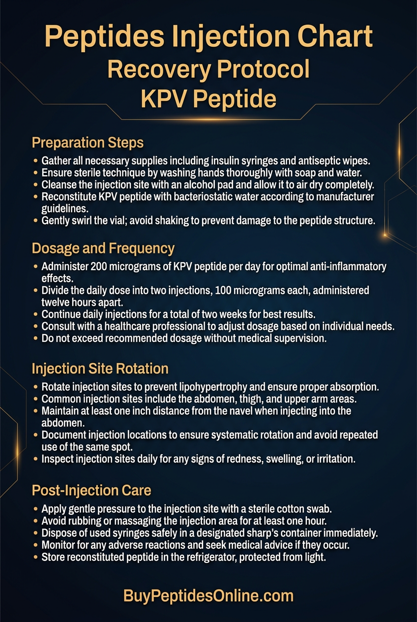
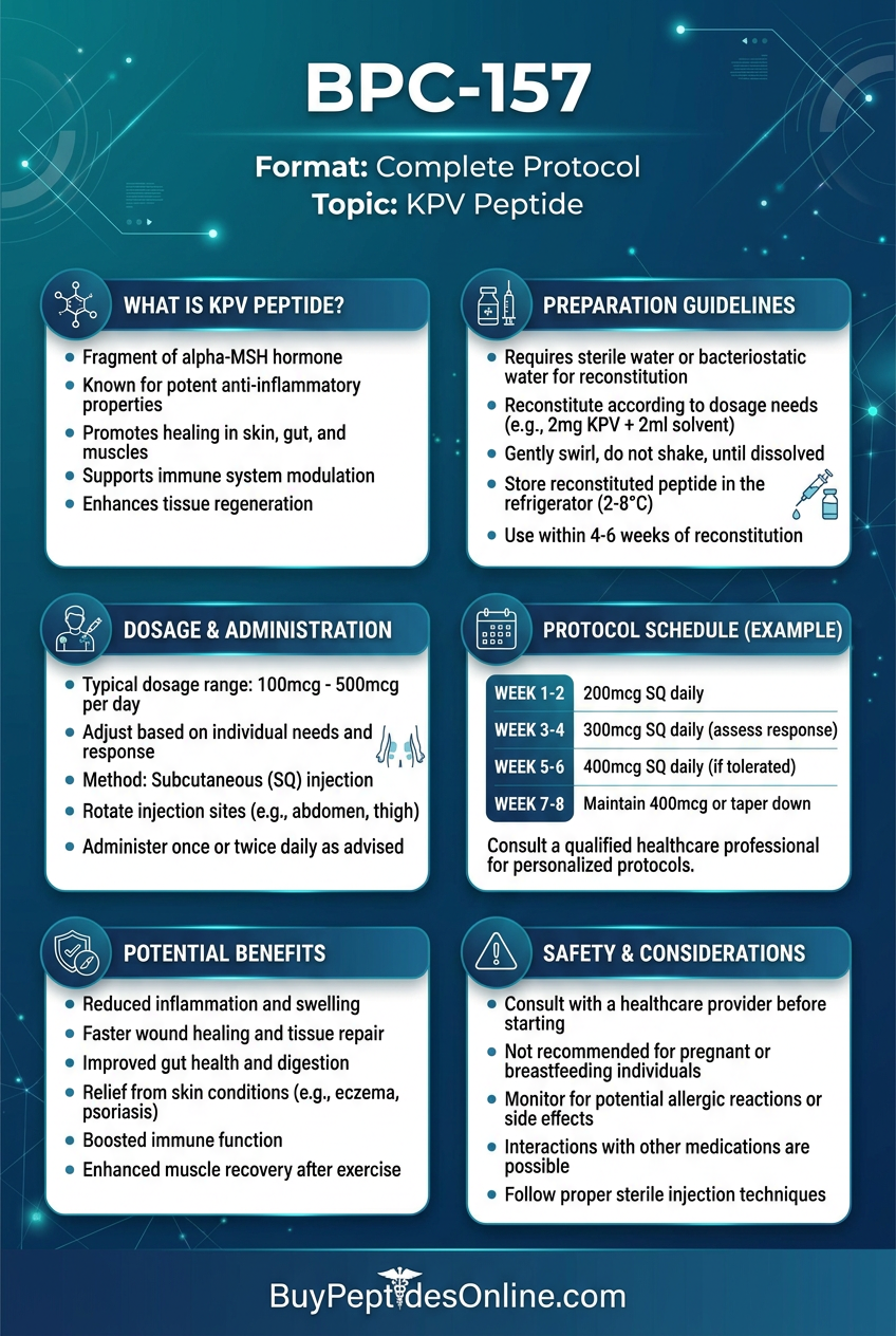
KPV is supplied as a lyophilized (freeze-dried) powder in research contexts. Once reconstituted in bacteriostatic water or sterile saline, it should be stored at 2-8°C (refrigerated) and used within a reasonable timeframe (typically 2-4 weeks for refrigerated solutions). For longer-term storage, the lyophilized powder should be kept at -20°C and protected from light and moisture. Repeated freeze-thaw cycles degrade the peptide and should be avoided.
KPV is generally considered chemically stable in its lyophilized form. Its small size and lack of disulfide bonds means it is less susceptible to some forms of degradation that affect larger, more structurally complex peptides.
---
Research Highlights: What the Studies Show
The research base for KPV spans multiple inflammatory conditions and delivery routes. While the majority of evidence comes from preclinical models, the consistency of findings across different tissues and experimental systems is notable.
Gastrointestinal Inflammation Research
The gut is where KPV research is most developed and most compelling. Inflammatory bowel disease—encompassing Crohn's disease and ulcerative colitis—represents a major unmet medical need, and the limitations of existing therapies (steroids, immunosuppressants, biologics) create genuine interest in alternatives.
The landmark 2018 mouse ulcerative colitis study remains the most-cited KPV study. Using a dextran sulfate sodium (DSS)-induced colitis model in mice (n=12/group), oral KPV at 2 mg/kg twice daily for 7 days produced a 72% reduction in intestinal inflammation compared to untreated controls. Histological scoring, cytokine measurements, and disease activity indices all showed improvement. Critically, this result was comparable to prednisolone (a standard-of-care corticosteroid) while avoiding the immunosuppressive and metabolic side effects associated with steroid use.
Further studies have examined KPV in nanoparticle-delivered forms, demonstrating enhanced colonic tissue concentrations and improved efficacy compared to free peptide oral administration. Nanoparticle-encapsulated KPV has shown preferential uptake by inflamed colonic epithelium and macrophages—a targeting mechanism that could theoretically reduce systemic exposure while maximizing local therapeutic effect.
Research has also examined KPV's effects on the gut microbiome in inflammation models. Some data suggest that by reducing inflammatory signaling in the gut epithelium, KPV may create conditions more favorable to commensal bacterial colonization—an indirect benefit for overall gut health that warrants further investigation.
Dermatological Research
KPV's skin research builds on the established role of α-MSH in cutaneous inflammation. The skin is rich in MC1R-expressing cells including keratinocytes, melanocytes, and dermal fibroblasts, making it a logical target for melanocortin-derived anti-inflammatory peptides.
In human keratinocyte cell culture studies, KPV at 10 μM applied for 48 hours reduced IL-8 production by 65%. IL-8 is a potent neutrophil chemoattractant that plays a central role in the inflammatory cascade of conditions like psoriasis, atopic dermatitis, and contact dermatitis. This reduction in IL-8 would theoretically reduce neutrophil infiltration and the associated tissue damage.
Additional keratinocyte studies have shown KPV reducing TNF-α-stimulated production of multiple pro-inflammatory mediators, with effects on both the NF-κB pathway and the MAPK (mitogen-activated protein kinase) cascade. The peptide has also demonstrated some ability to reduce UV-induced inflammatory responses in skin cells—a finding with potential relevance to photodermatitis and UV-related skin damage.
Topical formulation studies have examined KPV in cream and gel vehicles, confirming penetration through the stratum corneum and into the viable epidermis. Animal models of contact hypersensitivity have shown reduced ear swelling and inflammatory cell infiltration with topical KPV application, supporting its potential utility in allergic skin conditions.
Systemic Inflammation and Sepsis Research
In rat sepsis models (n=8/group), intravenous KPV at 1 mg/kg over 24 hours produced a 58% reduction in mortality rate compared to controls. This is a striking finding given that sepsis mortality remains a major clinical challenge, and it speaks to KPV's ability to modulate systemic inflammatory cascades when administered systemically.
The mechanism in sepsis models appears to involve suppression of the cytokine storm—the dysregulated, overwhelming inflammatory response that drives organ failure in severe sepsis. By dampening NF-κB-driven cytokine production across multiple cell types simultaneously, KPV may help contain the inflammatory cascade before it becomes irreversible.
Other systemic inflammation models have examined KPV in peritonitis, endotoxemia, and systemic lupus erythematosus-like conditions. While these studies are generally smaller and less replicated than the gut and skin work, they consistently support the conclusion that KPV has meaningful anti-inflammatory activity across multiple tissue compartments.
Summary Research Table
| Condition | Model | Dose | Duration | Key Result |
|---|---|---|---|---|
| Ulcerative colitis | Mice (n=12/group) | 2 mg/kg oral | 7 days | 72% ↓ inflammation vs control |
| Atopic/contact dermatitis | Human keratinocytes | 10 μM topical | 48h | IL-8 reduced by 65% |
| Sepsis | Rat (n=8/group) | 1 mg/kg IV | 24h | 58% ↓ mortality rate |
| Colitis (nanoparticle) | Mice | Encapsulated oral | 7-14 days | Enhanced tissue concentration vs free KPV |
| UV skin inflammation | Keratinocyte culture | 1-10 μM | 24-48h | Reduced TNF-α, IL-6 production |
| Peritonitis | Rodent models | 0.5-2 mg/kg IP | Acute | Reduced neutrophil infiltration |
---
Pharmacological Comparisons: KPV in Context
Understanding KPV's place in the research landscape requires comparing it to related compounds—both its parent molecule and other anti-inflammatory peptides that occupy similar research niches.
KPV vs. Full-Length α-MSH
**Alpha-MSH** is KPV's parent molecule and shares its anti-inflammatory mechanisms. The key differences are:
α-MSH: engages all five melanocortin receptors (MC1R-MC5R), including MC2R (ACTH receptor) and MC4R (appetite/sexual function). This produces a broader pharmacological profile with more potential off-target effects.
KPV: shows preferential activity at MC1R and MC3R with reduced engagement of MC4R, providing more targeted anti-inflammatory activity without the same degree of appetite suppression, sexual function effects, or pigmentation changes.
α-MSH: has greater potency at some receptor subtypes but carries the full hormonal activity of the intact neuropeptide.
KPV: offers a cleaner research tool for studying the anti-inflammatory arm of melanocortin signaling in isolation.
For gut-targeted research specifically, KPV's oral partial bioavailability gives it a practical advantage over α-MSH, which is more susceptible to gastrointestinal degradation.
KPV vs. BPC-157
BPC-157 is perhaps the most widely researched healing peptide in the research community, and it is frequently discussed alongside KPV for gut applications. The comparison is instructive:
BPC-157: (Body Protection Compound-157) is a 15-amino-acid peptide derived from a gastric protective protein. Its primary mechanisms involve angiogenesis stimulation, growth factor upregulation, and nitric oxide pathway modulation. It promotes tissue repair and regeneration.
KPV: works primarily through immune modulation—suppressing inflammatory signaling and expanding regulatory T cells. It does not have the same direct tissue-regenerative properties as BPC-157.
In gut healing research, the two peptides are complementary rather than redundant: KPV addresses the inflammatory component while BPC-157 promotes structural repair and regeneration.
This mechanistic complementarity is the scientific rationale for the KPV + BPC-157 stack discussed later in this article.
KPV vs. Thymosin Alpha-1
Thymosin Alpha-1 (TA1) is a 28-amino-acid peptide with broad immune-modulating properties. Its primary mechanism involves adaptive immunity—enhancing T cell differentiation, natural killer cell activity, and antigen presentation. It is used in research contexts for immune support, chronic viral infections, and cancer immunotherapy adjuncts.
KPV operates primarily on innate immunity—the rapid, non-specific first-line immune response mediated by macrophages, neutrophils, and dendritic cells. The two peptides therefore modulate different arms of the immune system, creating a rationale for combination research in conditions where both innate and adaptive immune dysregulation are present.
KPV vs. GHK-Cu
**GHK-Cu** (copper tripeptide-1) is another tripeptide with anti-inflammatory properties, particularly relevant in skin research. Like KPV, it is small enough for topical delivery. However, GHK-Cu's primary mechanisms involve copper-dependent enzyme activation, collagen synthesis stimulation, and antioxidant activity—a fundamentally different pharmacological profile from KPV's receptor-mediated immune modulation. In skin inflammation research, GHK-Cu may be more relevant for wound healing and structural repair, while KPV addresses the inflammatory component more directly.
KPV vs. LL-37
**LL-37** is a 37-amino-acid antimicrobial peptide (AMP) with both direct antimicrobial and immunomodulatory properties. It modulates innate immune responses, but its mechanisms involve direct membrane disruption of pathogens, TLR signaling modulation, and chemotaxis regulation—distinct from KPV's melanocortin receptor-mediated pathway. LL-37 is more relevant in contexts involving microbial infection alongside inflammation, while KPV is more specifically targeted at sterile or immune-mediated inflammation.
---
Research Applications: Who Is KPV Studied For?
Based on the existing preclinical literature, KPV research has been most concentrated in the following areas:
Inflammatory Bowel Disease Models
Crohn's disease and ulcerative colitis research represents the most developed application area for KPV. The combination of oral/local delivery potential, dual NF-κB/Treg mechanism, and demonstrated efficacy in DSS colitis models makes KPV particularly attractive for IBD research. The comparison to prednisolone in the 2018 study—same efficacy, different side effect profile—is the kind of finding that drives continued research interest.
Skin Inflammatory Conditions
Psoriasis, atopic dermatitis, and contact hypersensitivity models have all been examined with KPV. The peptide's topical deliverability (below 500 Da) and its activity on keratinocyte cytokine production make it a logical candidate for skin-targeted research. The IL-8 reduction finding in keratinocytes is particularly relevant to neutrophil-driven skin inflammation.
Systemic Inflammatory Conditions
The sepsis mortality data, while from a small animal study, has generated interest in KPV for systemic inflammatory conditions including sepsis, systemic inflammatory response syndrome (SIRS), and potentially autoimmune conditions with significant innate immune involvement.
Gut Permeability and Barrier Function
Beyond frank IBD, KPV's barrier-protective effects have attracted interest in the context of "leaky gut" research—conditions where intestinal permeability is increased without necessarily meeting diagnostic criteria for IBD. This is an active and somewhat controversial research area, but KPV's demonstrated effects on tight junction protein expression give it mechanistic relevance.
---
Practical Research Protocol Considerations
*The following reflects dosing and administration parameters used in preclinical research studies. These are not clinical dosing recommendations and should not be interpreted as medical advice.*
Research Dosing Overview
| Goal | Route | Dose | Frequency | Cycle |
|---|---|---|---|---|
| Gut inflammation | Oral | 1-2 mg/kg | 2x daily | 2-4 weeks |
| Skin conditions | Topical | 0.1-0.5% cream | Daily | As needed |
| Systemic support | SubQ | 100-200 mcg/kg | 3x weekly | 3 weeks |
| Acute systemic (research) | IV | 1 mg/kg | Single/acute | Per protocol |
Oral Administration Considerations
Oral KPV research protocols typically use doses in the 1-2 mg/kg range, administered twice daily to account for the relatively short half-life and the degradation that occurs during gastrointestinal transit. Timing relative to meals may influence absorption—some researchers prefer administration in a fasted state to reduce competition from dietary peptides for PepT1 transporter capacity.
Nanoparticle-encapsulated formulations, when available, may allow lower effective doses due to improved delivery efficiency. However, standard lyophilized KPV powder reconstituted in water is the most commonly used form in research settings.
Topical Formulation Considerations
For dermatological research applications, KPV is typically formulated in cream, gel, or serum vehicles at concentrations of 0.1-0.5% (1-5 mg/mL). The peptide's water solubility facilitates incorporation into aqueous-based formulations. Penetration enhancers (such as propylene glycol or certain surfactants) may be added to improve dermal delivery, though KPV's size already provides reasonable passive penetration.
Stability in topical formulations is a consideration—peptides can degrade in cream vehicles over time, particularly at elevated temperatures. Formulations should be stored refrigerated and used within the manufacturer's recommended timeframe.
Subcutaneous Administration
For systemic research applications, subcutaneous injection of reconstituted KPV is the most practical route. Reconstitution is typically performed with bacteriostatic water (for multi-use vials) or sterile water (for single-use). Standard peptide reconstitution practices apply: inject diluent slowly down the side of the vial, allow to dissolve without shaking, inspect for clarity before use.
Reconstitution and Handling
1. Allow the lyophilized vial to reach room temperature before opening to prevent moisture condensation
2. Use a clean, sterile technique throughout
3. Inject diluent slowly down the side of the vial—do not inject directly onto the powder
4. Gently swirl (do not shake) to dissolve
5. Inspect the solution: it should be clear and colorless
6. Label the vial with the date of reconstitution
7. Store reconstituted solution at 2-8°C; use within 2-4 weeks
8. For long-term storage of lyophilized powder: -20°C, protected from light and moisture
Timing and Cycling
Research protocols have used KPV in cycles of 2-4 weeks for gut applications, with some studies extending to 6 weeks without apparent adverse effects. The rationale for cycling (rather than continuous use) in research contexts is partly practical (to assess outcomes at defined endpoints) and partly precautionary (given limited long-term safety data). Continuous-use protocols have not been extensively studied.
---
Stacking Considerations: KPV in Combination Research
KPV's specific mechanism—immune modulation via melanocortin receptors—makes it mechanistically complementary to several other research peptides. The following combinations have scientific rationale based on non-overlapping or synergistic mechanisms.
KPV + BPC-157: The Gut Healing Stack
The most commonly discussed KPV combination is with BPC-157. The scientific rationale is elegant: in gut inflammation, two distinct processes need to be addressed—the inflammatory cascade that is damaging tissue, and the structural repair needed to restore mucosal integrity. KPV addresses the first component through NF-κB suppression and Treg expansion; BPC-157 addresses the second through angiogenesis stimulation, growth factor upregulation, and direct cytoprotective effects on gut epithelium.
This complementarity has been described as a "reduce and rebuild" strategy: KPV reduces the inflammatory environment that impedes healing, while BPC-157 actively stimulates the regenerative processes needed to restore tissue structure. Research into this combination is still largely theoretical in terms of formal co-administration studies, but the mechanistic logic is sound and the individual profiles of both peptides are well-characterized. Explore third-party tested BPC-157 from trusted suppliers for research use.
KPV + Thymosin Alpha-1: Innate + Adaptive Immune Modulation
Pairing KPV with Thymosin Alpha-1 creates a combination that addresses both the innate and adaptive arms of the immune system. KPV targets innate immune cells (macrophages, neutrophils, dendritic cells) through MC1R/MC3R signaling and NF-κB suppression; TA1 enhances adaptive immune function through T cell differentiation and NK cell activation. In conditions where both arms are dysregulated—as in many autoimmune conditions and chronic infections—this dual approach has theoretical appeal. Compare Thymosin Alpha-1 pricing from verified vendors for research procurement.
KPV + GHK-Cu: Comprehensive Skin Inflammation Protocol
For dermatological research, combining KPV's anti-inflammatory activity with GHK-Cu's collagen-stimulating and antioxidant properties creates a more comprehensive approach to skin inflammation. KPV suppresses the cytokine-mediated inflammatory cascade; GHK-Cu supports tissue repair and structural integrity. Both peptides are topically deliverable at their respective molecular weights, making co-formulation in a single topical vehicle feasible.
KPV + Selank: Stress-Inflammation Axis Research
There is growing research interest in the relationship between psychological stress and inflammatory disease—the so-called "stress-inflammation axis." Selank is an anxiolytic peptide with immunomodulatory properties that operates through different mechanisms (enkephalin-degrading enzyme inhibition, GABA modulation). In research contexts where stress-induced inflammation is a variable of interest, combining KPV's direct anti-inflammatory activity with Selank's stress-modulating properties could provide a more complete experimental model.
---
Safety Profile and Documented Side Effects
KPV's safety profile, as assessed across the available preclinical literature, is notably clean—though the caveat that long-term human safety data are limited must be clearly stated.
Acute Toxicity
No serious adverse events have been reported in published KPV studies across oral, topical, intravenous, and intraperitoneal administration routes. Acute toxicity studies in rodents have not identified a maximum tolerated dose at pharmacologically relevant doses. The peptide does not appear to cause local injection site reactions beyond those expected with any injectable compound.
Immunosuppression Risk
The most theoretically significant safety concern with KPV is the risk of excessive immunosuppression with prolonged high-dose use. Since KPV suppresses pro-inflammatory cytokine production and expands regulatory T cells, there is a theoretical possibility that very high doses or very long treatment durations could impair appropriate immune responses to infection or malignancy.
However, it is important to contextualize this risk. The doses used in research studies produce targeted modulation of dysregulated inflammation rather than global immune suppression. Unlike corticosteroids, which broadly suppress immune function through glucocorticoid receptor activation across virtually all immune cell types, KPV's mechanism is more targeted. The Treg-expanding effect is arguably immunoregulatory rather than immunosuppressive—it restores appropriate immune balance rather than simply suppressing immune activity.
That said, caution is warranted in research contexts involving subjects with active infections, immunodeficiency, or known malignancies. Concurrent use with other immunosuppressive agents is a theoretical concern that has not been formally studied.
Theoretical Hormonal Concerns
Given that KPV is derived from α-MSH and engages melanocortin receptors, there is a theoretical question about whether it could influence pigmentation, appetite, or other MC receptor-mediated functions with prolonged use. The available evidence suggests that KPV's receptor binding profile does not produce these effects at research doses—the peptide's selectivity for MC1R/MC3R anti-inflammatory signaling over MC4R-mediated appetite effects appears to hold in practice. However, this has not been formally assessed in long-term human studies.
Contraindications and Precautions
Based on theoretical considerations and the available preclinical literature:
Concurrent immunosuppressive drugs: Theoretical additive immunosuppression risk; not formally studied
Active serious infections: Theoretical concern about impaired immune response; use with caution in research protocols
Pregnancy and lactation: No data available; standard precautionary exclusion applies
Known malignancy: Theoretical concern about Treg expansion potentially reducing anti-tumor immune surveillance; not formally assessed
What Has Not Been Observed
Notably absent from the KPV safety literature are:
Hepatotoxicity
Nephrotoxicity
Cardiovascular effects
Endocrine disruption (at research doses)
Anaphylaxis or serious allergic reactions
Neurotoxicity
This clean safety signal, while encouraging, must be interpreted in the context of relatively small study sizes and limited human data.
---
Common Research Mistakes and Pitfalls
Researchers and research coordinators working with KPV should be aware of several common errors that can compromise experimental validity or reduce observed efficacy.
Mistake 1: Assuming All Peptide Vendors Supply Equivalent Quality
KPV's small size makes it relatively straightforward to synthesize, but this also means quality varies significantly between suppliers. Impurities in the form of truncated sequences, oxidized amino acids (particularly the valine residue), or residual synthesis reagents can reduce potency and introduce confounding variables. Research-grade KPV should come with HPLC purity data (ideally ≥98%) and mass spectrometry confirmation of molecular weight. Suppliers who cannot provide these certificates of analysis should be avoided for serious research applications.
Mistake 2: Inadequate Storage of Reconstituted Peptide
KPV in solution is more vulnerable to degradation than the lyophilized powder. Leaving reconstituted KPV at room temperature, exposing it to repeated light cycles, or allowing it to freeze and thaw multiple times will reduce active peptide concentration and compromise experimental reproducibility. Strict cold chain management is essential.
Mistake 3: Expecting Oral KPV to Have Systemic Effects
While KPV has meaningful oral bioavailability for gut-targeted applications, researchers should not assume that oral administration produces significant systemic blood levels sufficient for non-gastrointestinal endpoints. For systemic anti-inflammatory research applications, subcutaneous or intravenous routes are more appropriate. Oral administration is specifically suited to intestinal inflammation research.
Mistake 4: Using Single-Dose Protocols for Chronic Inflammation Models
KPV's short half-life means that single-dose or infrequent dosing is unlikely to maintain sufficient receptor engagement for chronic inflammation models. Research protocols for ongoing inflammatory conditions should use multiple daily doses or consider sustained-release formulation approaches.
Mistake 5: Overlooking the Nanoparticle Literature
A significant body of KPV research uses nanoparticle-encapsulated formulations that produce substantially different pharmacokinetics and tissue distribution compared to free peptide. Researchers comparing their results to published studies should carefully note whether the reference study used free KPV or nanoparticle-delivered KPV, as the two are not directly comparable in terms of effective dose.
Mistake 6: Ignoring the Treg Component in Outcome Assessment
Many researchers focus exclusively on cytokine levels (TNF-α, IL-6, IL-1β) as outcome measures for KPV studies, overlooking the Foxp3+ Treg expansion that is a core part of KPV's mechanism. Comprehensive assessment of KPV's effects should include flow cytometric analysis of Treg populations in relevant lymphoid tissues, particularly for gut and systemic inflammation models.
---
Research Outlook: Where Is KPV Science Headed?
KPV occupies an interesting position in the peptide research landscape: well-characterized mechanistically, with consistent preclinical efficacy across multiple models, but with a significant gap between preclinical evidence and human clinical data. Several directions are shaping the future of KPV research.
Nanoparticle Delivery Systems
The development of increasingly sophisticated nanoparticle delivery systems for KPV represents perhaps the most active area of translational research. Hydrogel nanoparticles, lipid nanoparticles, and polymeric nanocarriers have all been explored as vehicles for oral KPV delivery to inflamed gut tissue. The goal is to achieve higher local concentrations in inflamed tissue while minimizing systemic exposure—a targeting strategy that would improve both efficacy and the safety profile.
Recent work has explored colitis-targeting nanoparticles that preferentially accumulate in inflamed colonic tissue based on the altered pH and enzymatic environment of inflamed mucosa. This kind of disease-state-specific targeting could represent a significant advance in KPV delivery for IBD applications.
Combination Biologics Research
There is growing interest in combining KPV with existing biologic therapies for IBD (such as anti-TNF antibodies or integrin inhibitors) to explore whether the combination produces additive or synergistic effects. The mechanistic rationale is that biologics typically block a single cytokine or adhesion molecule, while KPV's upstream NF-κB suppression affects multiple inflammatory mediators simultaneously—potentially providing broader coverage.
Skin Microbiome Interactions
Emerging research on the skin microbiome and its relationship to inflammatory skin conditions has created new questions about how KPV's anti-inflammatory effects interact with cutaneous microbial communities. Since skin inflammation significantly alters the microbiome composition (and vice versa), understanding whether KPV's anti-inflammatory effects have secondary benefits for microbiome balance is an active area of inquiry.
Neuroinflammation Applications
While less developed than the gut and skin literature, there is preliminary interest in KPV's potential for neuroinflammation research. MC1R and MC3R are expressed in the central nervous system, and neuroinflammation (mediated by activated microglia and astrocytes) shares mechanistic features with peripheral inflammation. Given the blood-brain barrier challenges for most peptides, the small size of KPV—which might facilitate CNS penetration—makes this a theoretically interesting direction.
Human Clinical Data
The most significant gap in the KPV literature is the absence of controlled human clinical trials. The preclinical data are compelling and consistent, but translation to human biology is never guaranteed. The safety profile and mechanistic rationale support the case for human trials, and it is likely that IBD will be the first indication pursued given the strength of the gut inflammation data. The development of nanoparticle delivery systems that can achieve reliable oral bioavailability will be a key enabling step for human gut-targeted trials.
---
KPV in the Broader Anti-Inflammatory Peptide Landscape
For researchers interested in the full spectrum of anti-inflammatory peptides, KPV occupies a specific and distinctive niche. The best anti-inflammatory peptides available for research cover a wide range of mechanisms, and understanding where KPV fits requires appreciating what makes it unique.
Unlike corticotropin-releasing hormone analogs that work through the HPA axis, or VIP (vasoactive intestinal peptide) that modulates inflammation through cAMP-dependent mechanisms in a broader neuroimmune context, KPV's melanocortin receptor-mediated mechanism is relatively specific and well-characterized. Unlike larger anti-inflammatory peptides that require injectable administration, KPV's oral and topical deliverability expands its practical research utility.
For researchers focused specifically on gut healing, the combination of KPV with BPC-157 represents the most mechanistically complete approach currently available in the research peptide space—addressing inflammation, barrier function, and tissue regeneration through complementary mechanisms. Detailed exploration of this combination is covered in the BPC-157 gut healing guide.
For researchers focused on skin inflammation, KPV's combination with GHK-Cu—the copper tripeptide with established collagen-stimulating properties—creates a comprehensive approach to cutaneous inflammatory conditions. Both peptides are topically deliverable and mechanistically complementary.
---
Frequently Asked Questions
### What exactly is KPV and where does it come from?
KPV (Lysine-Proline-Valine) is a synthetic tripeptide that replicates the C-terminal three amino acids of alpha-melanocyte-stimulating hormone (α-MSH), a naturally occurring neuropeptide produced by the pituitary gland and peripheral tissues. Researchers studying α-MSH's anti-inflammatory properties in the 1990s identified the C-terminal KPV sequence as the minimal fragment responsible for those effects. At just 391 Daltons and three amino acids, it is one of the smallest bioactive peptides studied for anti-inflammatory applications. In research contexts, it is produced through chemical peptide synthesis and supplied as a lyophilized powder.
### How does KPV differ from taking alpha-MSH directly?
Full-length alpha-MSH engages all five melanocortin receptor subtypes (MC1R through MC5R), producing effects that include pigmentation changes (via MC1R), adrenal stimulation (via MC2R), appetite suppression and sexual function effects (via MC4R), and exocrine gland regulation (via MC5R). KPV preferentially engages MC1R and MC3R in ways that activate anti-inflammatory signaling without the same degree of engagement with MC4R-mediated pathways. The result is that KPV retains the anti-inflammatory properties of α-MSH while avoiding the hormonal, pigmentation, and appetite effects of the full molecule. For researchers specifically interested in inflammation modulation, this makes KPV a cleaner and more targeted research tool.
### Can KPV actually be taken orally, and does it work that way?
Yes, with important qualifications. KPV's three-amino-acid structure makes it a substrate for PepT1, the intestinal di/tripeptide transporter, allowing partial absorption across the gut epithelium. This is unusual among research peptides, most of which are rapidly degraded in the gastrointestinal tract. The 2018 mouse colitis study demonstrating 72% inflammation reduction used oral administration, validating this route for gut-targeted applications. However, oral KPV likely produces primarily local gut effects rather than significant systemic blood levels. For systemic anti-inflammatory research applications, subcutaneous or intravenous routes are more appropriate. Nanoparticle encapsulation can improve oral delivery efficiency and tissue targeting.
### What are the most important safety considerations for KPV research?
The preclinical safety profile of KPV is notably clean—no serious adverse events have been reported across multiple administration routes in published studies. The primary theoretical safety concern is excessive immunosuppression with very high doses or prolonged use, given KPV's activity in suppressing pro-inflammatory cytokines and expanding regulatory T cells. In practice, the doses used in research studies appear to produce targeted immunomodulation rather than global immune suppression. Concurrent use with other immunosuppressive agents is a theoretical concern. Research protocols should exclude subjects with active serious infections. Human long-term safety data are limited, and this represents the most significant gap in the current evidence base.
### What makes KPV particularly interesting for gut inflammation research?
Several converging factors make KPV especially relevant for gut inflammation research. First, it has demonstrated oral/local bioavailability that allows it to reach inflamed gut tissue without requiring injection—unusual among research peptides. Second, its dual mechanism (NF-κB suppression + Treg expansion) addresses both the acute inflammatory response and the underlying immune dysregulation that perpetuates chronic gut inflammation. Third, the 2018 mouse colitis study showing efficacy comparable to prednisolone without corticosteroid side effects is a compelling proof-of-concept. Fourth, nanoparticle delivery systems under development may allow targeted delivery to inflamed colonic tissue with minimal systemic exposure—a particularly attractive profile for IBD research.
### How does KPV compare to BPC-157 for gut healing research?
BPC-157 and KPV are mechanistically complementary rather than comparable. BPC-157 works primarily through angiogenesis stimulation, growth factor upregulation, and direct cytoprotective effects—it promotes tissue repair and regeneration. KPV works through immune modulation—suppressing inflammatory signaling and expanding regulatory T cells. In gut inflammation research, BPC-157 is better suited to the repair and regeneration phase, while KPV addresses the inflammatory cascade that causes and perpetuates tissue damage. The combination is sometimes described as "reduce and rebuild"—KPV reduces inflammation, BPC-157 rebuilds tissue. For comprehensive gut inflammation research, the combination has more complete mechanistic coverage than either peptide alone.
### What does the research say about KPV for skin conditions?
KPV's skin research is primarily based on cell culture studies using human keratinocytes, with supporting data from animal contact hypersensitivity models. In keratinocyte studies, KPV at 10 μM reduced IL-8 production by 65% over 48 hours—a meaningful reduction in a key neutrophil-attracting cytokine central to skin inflammatory conditions. Animal models of contact hypersensitivity have shown reduced inflammatory cell infiltration with topical KPV application. KPV's molecular weight (391 Da) is below the 500 Dalton threshold for passive skin penetration, making topical delivery feasible without specialized carriers. Human clinical data for skin conditions are not yet available, and the existing evidence base—while mechanistically compelling—is primarily preclinical.
---
Key Takeaways
KPV is the smallest clinically studied anti-inflammatory peptide: , at just three amino acids and 391 Daltons—yet its mechanism is sophisticated, targeting NF-κB suppression and Treg expansion simultaneously
Derived from α-MSH: , it retains the parent molecule's anti-inflammatory properties without the hormonal, pigmentation, and appetite effects of the full 13-amino-acid sequence
Oral and topical bioavailability: are genuine advantages over larger peptides, making KPV uniquely suited for gut-targeted and dermatological research applications
The 2018 colitis mouse study: remains the landmark finding: 72% reduction in intestinal inflammation comparable to prednisolone, without immunosuppressive side effects
Mechanistically: , KPV works through MC1R/MC3R binding → NF-κB suppression → reduced TNF-α, IL-6, IL-1β, IL-8; and simultaneously through Foxp3+ Treg expansion in gut-associated lymphoid tissue
Research-grade KPV: should come with HPLC purity certificates (≥98%) and mass spectrometry confirmation—quality varies significantly between suppliers; lab-tested KPV is available from verified research suppliers
Pairs powerfully with BPC-157: for comprehensive gut healing research (inflammation reduction + tissue regeneration), and with Thymosin Alpha-1 for broader immune modulation research
Human safety data are limited but promising: —no serious adverse events in preclinical studies, but long-term human trials are needed before clinical conclusions can be drawn
Nanoparticle delivery systems: represent the most exciting translational frontier, potentially enabling targeted delivery to inflamed gut tissue with minimal systemic exposure
All information here is for educational and research purposes only; KPV is not an approved drug and this does not constitute medical advice
---
Continue Your Peptide Research
🔬 Explore our peptide database — Browse 500+ research peptide profiles with mechanisms of action, dosing protocols, and clinical evidence summaries.
🤖 Have questions? — Ask PeptideAI, our research assistant, for personalized peptide guidance based on the latest studies.
📚 Want more guides? — Browse all research articles covering peptide science, comparisons, and buying guides.
Related Articles on BuyPeptidesOnline.com
Continue your research with these in-depth guides: