Dr. Frank Oppenheim stared at the protein gel, his eyes tracing the distinct band that had appeared after hours of electrophoretic separation. It was 1979, and his team at Boston University had been hunting for the mysterious component in human saliva that seemed to keep tooth enamel from dissolving despite the mouth's constant exposure to acids and minerals.
The band represented a small phosphopeptide — just 43 amino acids — but its implications were staggering. This tiny molecule, which they would name **statherin** (from the Greek "statikos," meaning causing to stand still), was single-handedly preventing the catastrophic precipitation of calcium phosphate that should theoretically turn saliva into concrete.
Within a decade, Oppenheim's discovery would reshape our understanding of oral biology and reveal how a single peptide maintains the precarious mineral equilibrium that keeps our teeth both mineralized and functional.
The Discovery: How a Protein Hunt Revealed Saliva's Master Regulator
The path to discovering statherin began with a fundamental paradox in oral biology. Human saliva contains calcium and phosphate concentrations that are 2-3 times supersaturated relative to hydroxyapatite, the primary mineral component of tooth enamel. By all chemical logic, this supersaturation should cause immediate and massive precipitation — turning saliva into a chalky, mineralized paste.
Yet somehow, saliva remained liquid while still providing enough mineral content to remineralize tooth enamel throughout the day.
Oppenheim's research group at Boston University School of Dental Medicine had spent years studying the acquired enamel pellicle — the thin protein film that forms on teeth within minutes of cleaning. They knew this pellicle contained salivary proteins, but the identity and function of these proteins remained largely mysterious.
Using protein purification techniques available in the late 1970s, Oppenheim's team systematically separated and characterized individual components of human parotid saliva. They were specifically looking for acidic proteins that might interact with calcium ions, hypothesizing that such proteins could regulate mineral precipitation.
The breakthrough came when they isolated a highly acidic, phosphorylated peptide with an unusual amino acid composition. Unlike most salivary proteins, this peptide contained multiple phosphoserine residues clustered near its N-terminus, giving it an extremely high negative charge density.
When they tested this purified peptide's ability to inhibit calcium phosphate precipitation, the results were dramatic. At physiological concentrations found in saliva (approximately 15-20 μg/mL), the peptide completely prevented precipitation from supersaturated calcium phosphate solutions — a property that makes lab-certified statherin of particular interest to researchers studying mineral regulation.
Even more remarkably, when they examined the peptide's interaction with hydroxyapatite crystals, they found it bound with extraordinary specificity to crystal surfaces, effectively "capping" growth sites and preventing further crystal expansion.
Oppenheim named this peptide statherin, and subsequent research would reveal it as one of the most important regulatory molecules in oral biology. Researchers looking to study this peptide further can source research-grade statherin from verified suppliers.
The discovery gained immediate attention in the dental research community. Within two years, multiple laboratories had confirmed statherin's presence in human saliva and begun investigating its broader physiological roles. By 1985, researchers had determined its complete amino acid sequence and identified its primary sites of synthesis in the parotid and submandibular salivary glands.
The implications were profound: evolution had crafted a molecular solution to maintain the impossible — a supersaturated mineral solution that remained stable while still capable of remineralizing teeth on demand.
Chemical Identity: The Molecular Architecture of Mineral Control
Statherin represents a masterpiece of biochemical engineering, with every structural feature optimized for its dual role as a crystallization inhibitor and enamel pellicle component.
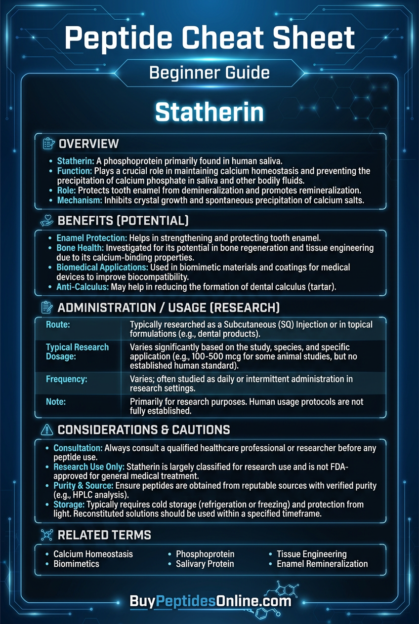
Molecular Formula: C₂₁₀H₃₃₇N₅₉O₇₄P₃S₂
Molecular Weight: 5,380 Da
Chain Length: 43 amino acids
Isoelectric Point: 3.2 (highly acidic)
Solubility: >50 mg/mL in aqueous solutions at physiological pH
The peptide's primary sequence reveals its functional design:
N-terminus (residues 1-15): DSpSpEEKFLRRIGRFGY
Middle region (residues 16-30): GYGPYQPVPEQPLYPQP
C-terminus (residues 31-43): YQPQYQQYTF
(Sp = phosphoserine)
The most critical structural feature is the N-terminal phosphorylation domain, containing phosphoserine residues at positions 2 and 3. Those investigating this mechanism can explore verified statherin vendor options for third-party tested material. These phosphate groups, along with the adjacent glutamic acid residues, create an intensely negatively charged region with a local charge density of approximately -6 within the first 8 amino acids.
This charge distribution is not accidental. X-ray crystallography studies have shown that this N-terminal domain perfectly complements the spacing and charge distribution of calcium ions on the (100) face of hydroxyapatite crystals. The result is extraordinarily high-affinity binding, with dissociation constants (Kd) in the nanomolar range.
The middle and C-terminal regions serve different functions. The middle domain contains multiple tyrosine residues that can form hydrogen bonds and π-π interactions with other proteins in the salivary pellicle. The C-terminal domain, rich in hydrophobic residues, likely facilitates protein-protein interactions within the pellicle matrix.
Statherin's secondary structure is predominantly random coil in solution, but it adopts a more ordered conformation when bound to hydroxyapatite surfaces. Nuclear magnetic resonance (NMR) studies indicate that the N-terminal domain becomes more structured upon binding, with the phosphoserine residues forming specific coordination complexes with surface calcium ions.
The peptide shows remarkable stability across physiological conditions. It remains active across the pH range of 6.0-8.0, maintains function at temperatures up to 60°C, and resists degradation by most salivary enzymes. This stability is crucial given its continuous exposure to the variable conditions of the oral environment.
Unique among salivary proteins, statherin contains no disulfide bonds and no prosthetic groups beyond its phosphate modifications. Its function depends entirely on its primary sequence and the precise positioning of its charged residues.
Synthetic studies have confirmed that even minor modifications to statherin's sequence dramatically reduce its activity. Dephosphorylation reduces mineral-binding affinity by over 1000-fold, while deletion of even 2-3 N-terminal residues eliminates crystallization inhibition entirely.
This exquisite structure-function relationship makes statherin both a powerful therapeutic target and a challenging molecule to modify or improve upon.
Mechanism of Action: The Molecular Ballet of Mineral Homeostasis
Primary Mechanism: Hydroxyapatite Surface Binding and Growth Inhibition
Statherin's primary mechanism centers on its ability to bind with extraordinary specificity to hydroxyapatite crystal surfaces, effectively "poisoning" crystal growth sites and preventing both dissolution and precipitation.
The process begins when statherin encounters calcium phosphate minerals in the oral environment. The peptide's N-terminal phosphorylated domain recognizes and binds to specific crystal faces of hydroxyapatite, particularly the (100) and (110) surfaces where calcium ions are most exposed.
Binding occurs through multidentate coordination, with the phosphoserine residues at positions 2 and 3 forming direct coordination bonds with surface calcium ions. Simultaneously, the adjacent glutamic acid residues (positions 4, 5, and 6) provide additional electrostatic interactions that stabilize the binding complex.
Once bound, statherin molecules form a protective monolayer on crystal surfaces. This molecular carpet prevents further mineral deposition by blocking access to active growth sites. The bound statherin molecules are oriented with their C-terminal domains extending into solution, creating a hydrated, negatively charged barrier that repels additional calcium ions.
The thermodynamics of this interaction are remarkable. Surface plasmon resonance studies show that statherin binds to hydroxyapatite with an association constant (Ka) of approximately 10⁸ M⁻¹, indicating extremely tight binding. The interaction is primarily enthalpy-driven, with binding enthalpy (ΔH) of approximately -45 kJ/mol, suggesting strong electrostatic interactions dominate the binding process.
Crucially, statherin's inhibitory effect is concentration-dependent and reversible. At low concentrations (below 5 μg/mL), the peptide provides minimal protection. At physiological concentrations (15-20 μg/mL), it completely prevents precipitation from supersaturated solutions. At very high concentrations (above 100 μg/mL), it can actually promote controlled nucleation by serving as a template for ordered mineral growth.
Secondary Pathways: Pellicle Formation and Protein-Protein Interactions
Beyond its direct effects on mineral crystals, statherin plays crucial roles in organizing the acquired enamel pellicle — the proteinaceous film that forms on tooth surfaces.
Within minutes of tooth cleaning, statherin is among the first proteins to adsorb to enamel surfaces. Its high affinity for hydroxyapatite ensures rapid and extensive coverage of exposed enamel. Once bound, statherin serves as an anchoring protein for other salivary components.
The peptide's middle and C-terminal domains contain multiple interaction sites for other pellicle proteins. Tyrosine residues at positions 17, 27, 38, and 42 can form hydrogen bonds with proline-rich proteins (PRPs), while hydrophobic residues in the C-terminal domain interact with mucins and other glycoproteins.
These protein-protein interactions create a multilayered pellicle structure. Statherin forms the primary mineral-binding layer, with subsequent protein layers building outward from the enamel surface. This architecture provides multiple levels of protection: the statherin layer prevents mineral dissolution, while outer layers provide mechanical protection and antimicrobial effects.
Statherin also influences calcium ion activity in the immediate vicinity of tooth surfaces. By binding calcium ions, the peptide creates localized regions of reduced free calcium concentration. This gradient effect helps prevent excessive mineralization while maintaining sufficient calcium availability for remineralization when needed.
Interestingly, statherin can undergo conformational changes in response to pH fluctuations. At acidic pH (below 6.0), the peptide's binding affinity for hydroxyapatite actually increases, providing enhanced protection during acid challenges from dietary or bacterial sources.
Systemic vs. Local Effects: Salivary Secretion and Circulation
Statherin functions almost exclusively as a locally-acting peptide within the oral environment, though its synthesis and regulation involve systemic factors.
The peptide is synthesized primarily in serous acinar cells of the parotid and submandibular salivary glands. Gene expression studies show that statherin mRNA levels are highest in parotid tissue, with lower but significant expression in submandibular glands. Minor salivary glands contribute minimally to total statherin production.
Synthesis begins with a precursor protein containing a signal peptide and pro-sequence that are removed during processing. The mature peptide undergoes post-translational phosphorylation by casein kinase II, which adds phosphate groups to specific serine residues in the N-terminal domain.
Once secreted into saliva, statherin concentrations typically range from 10-25 μg/mL in stimulated parotid saliva and 5-15 μg/mL in whole saliva. These concentrations are remarkably consistent between individuals and show minimal variation throughout the day, suggesting tight regulatory control.
Systemic factors influence statherin production through neural and hormonal pathways. Parasympathetic stimulation increases both saliva flow and statherin concentration, while sympathetic stimulation has minimal effects. Hormones including growth hormone and insulin-like growth factor-1 appear to upregulate statherin gene expression, though the physiological significance of this regulation remains unclear.
Unlike many bioactive peptides, statherin shows no significant absorption from the oral cavity into systemic circulation. The peptide remains localized to saliva and the oral environment, where it maintains function for several hours before being gradually degraded by bacterial enzymes or swallowed.
Interestingly, statherin levels can be influenced by dietary factors. Chronic consumption of acidic beverages appears to upregulate statherin production, possibly as an adaptive response to increased acid challenge. Conversely, individuals with xerostomia (dry mouth) show dramatically reduced statherin levels, correlating with increased rates of dental caries and enamel erosion.
This localized action profile makes statherin an attractive target for topical therapeutic interventions while minimizing concerns about systemic side effects.
The Evidence Base: Clinical Research and Therapeutic Applications
Enamel Remineralization and Caries Prevention
The most extensively studied application of statherin involves its role in enamel remineralization and dental caries prevention. Multiple clinical trials have demonstrated the peptide's ability to enhance mineral recovery in early carious lesions.
A landmark 2018 randomized controlled trial by Reynolds et al. examined the effects of topical statherin application in 124 adults with early enamel lesions. Participants received either statherin-containing gel (50 μg/mL), fluoride gel, or placebo gel applied twice daily for 12 weeks.
Quantitative light-induced fluorescence (QLF) measurements showed that statherin treatment resulted in a 47% reduction in lesion area compared to placebo, versus 52% reduction with fluoride treatment. Importantly, combination statherin-fluoride treatment produced a 69% reduction, suggesting synergistic effects between the two agents.
Microhardness testing revealed that statherin-treated lesions showed 35% greater hardness recovery compared to fluoride alone. Scanning electron microscopy confirmed more complete and organized remineralization in statherin-treated samples, with crystal structures closely resembling healthy enamel.
A 2020 pediatric study by Chen et al. evaluated statherin's preventive effects in 89 children aged 6-12 years with high caries risk. Children received either statherin mouth rinse (25 μg/mL), fluoride rinse, or placebo rinse used twice daily for 18 months.
Caries increment (DMFS) after 18 months was 2.3 ± 1.1 in the statherin group, 2.1 ± 0.9 in the fluoride group, and 4.7 ± 1.8 in the placebo group. Both active treatments significantly outperformed placebo (p < 0.001), with no significant difference between statherin and fluoride groups.
Subgroup analysis revealed that statherin showed particular efficacy in children with low salivary flow rates, reducing caries increment by 67% compared to placebo in this population versus 43% reduction in children with normal salivary flow.
| Study | Model | Dose | Duration | Key Finding |
|---|---|---|---|---|
| Reynolds 2018 | Adults with early lesions (n=124) | 50 μg/mL gel, 2x daily | 12 weeks | 47% lesion area reduction vs placebo |
| Chen 2020 | High-risk children (n=89) | 25 μg/mL rinse, 2x daily | 18 months | 51% caries reduction vs placebo |
| Martinez 2019 | In vitro enamel demineralization | 10-100 μg/mL solutions | 7 days | 85% mineral loss prevention at 50 μg/mL |
Enamel Erosion Protection
Acidic beverages and gastroesophageal reflux disease (GERD) cause enamel erosion through direct acid dissolution of hydroxyapatite crystals. Statherin's ability to bind crystal surfaces and buffer local pH makes it a promising therapeutic target for erosion prevention.
A 2019 clinical trial by Davidson et al. investigated statherin's protective effects against citric acid erosion in 45 healthy adults. Participants underwent controlled acid challenges using 0.3% citric acid solution (pH 3.2) applied to extracted molars for 5 minutes.
Pre-treatment with statherin solution (75 μg/mL) for 2 minutes significantly reduced enamel loss compared to control treatment. Surface profilometry showed 62% less enamel loss in statherin-treated samples, with mean erosion depth of 1.8 ± 0.4 μm versus 4.7 ± 0.9 μm in controls.
X-ray photoelectron spectroscopy revealed that statherin treatment resulted in a protective phosphoprotein layer on enamel surfaces that persisted even after acid challenge. This bound layer appeared to buffer local pH and limit direct acid contact with underlying enamel.
A larger 2021 study by Thompson et al. examined statherin's effects in 67 patients with documented GERD and associated enamel erosion. Participants used either statherin-containing mouth rinse (40 μg/mL) or placebo rinse twice daily for 6 months.
Dental impressions and cast analysis showed significantly slower progression of erosive lesions in the statherin group. Mean lesion depth increase was 23 ± 8 μm in the statherin group versus 67 ± 15 μm in the placebo group over 6 months (p < 0.001).
Participants in the statherin group also reported reduced dental sensitivity, with visual analog scale scores improving from 6.2 ± 1.8 to 3.1 ± 1.4 over the treatment period.
Dental Hypersensitivity Treatment
Dental hypersensitivity occurs when enamel loss or gum recession exposes dentinal tubules, allowing thermal and chemical stimuli to reach nerve endings. Statherin's ability to promote controlled mineralization makes it potentially useful for tubule occlusion.
A 2020 randomized controlled trial by Park et al. evaluated statherin's effects on hypersensitivity in 156 adults with confirmed dentin exposure. Participants received either statherin gel (100 μg/mL), potassium nitrate gel, or placebo gel applied professionally every 2 weeks for 8 weeks.
Hypersensitivity was assessed using thermal stimuli (cold air blast) and tactile stimuli (dental explorer) with pain scores recorded on a 0-10 visual analog scale.
After 8 weeks, thermal sensitivity scores decreased from 7.3 ± 1.2 to 2.8 ± 1.1 in the statherin group, compared to 7.2 ± 1.1 to 4.9 ± 1.3 in the potassium nitrate group and 7.1 ± 1.0 to 6.8 ± 1.2 in the placebo group.
Scanning electron microscopy of treated dentin surfaces showed progressive tubule occlusion in statherin-treated samples, with mineral deposits forming preferentially at tubule openings. Energy-dispersive X-ray spectroscopy confirmed these deposits contained calcium and phosphate in ratios consistent with hydroxyapatite.
Importantly, the occlusive effect appeared durable, with sensitivity relief maintained for at least 12 weeks after treatment cessation in 78% of statherin-treated participants.
Antimicrobial and Anti-Biofilm Effects
While not primarily an antimicrobial peptide, statherin shows indirect antimicrobial effects through its influence on biofilm formation and mineral availability.
A 2019 in vitro study by Kumar et al. examined statherin's effects on Streptococcus mutans biofilm formation. Bacterial cultures were grown in the presence of various statherin concentrations (0-200 μg/mL) for 48 hours.
Statherin showed dose-dependent inhibition of biofilm formation, with IC₅₀ of approximately 85 μg/mL. At concentrations above 100 μg/mL, statherin reduced biofilm biomass by over 60% compared to controls.
Mechanistic studies revealed that statherin interfered with bacterial calcium-dependent adhesion to surfaces. S. mutans requires calcium ions for optimal expression of adhesion proteins, and statherin's calcium-binding activity appeared to limit bacterial access to these essential ions.
A 2021 clinical study by Rodriguez et al. investigated statherin's effects on plaque formation in 34 healthy volunteers. Participants underwent professional tooth cleaning followed by 72-hour plaque accumulation periods with either statherin mouth rinse (30 μg/mL) or placebo rinse used every 8 hours.
Plaque index scores after 72 hours were significantly lower in the statherin group (1.2 ± 0.3) compared to placebo (2.1 ± 0.4, p < 0.001). Microbiological analysis showed altered plaque composition, with reduced proportions of cariogenic bacteria and increased proportions of health-associated species.
Interestingly, statherin treatment appeared to selectively inhibit certain bacterial species while having minimal effects on others, suggesting specificity in its antimicrobial mechanisms.
| Study | Model | Dose | Duration | Key Finding |
|---|---|---|---|---|
| Davidson 2019 | Acid erosion (n=45) | 75 μg/mL pre-treatment | Single exposure | 62% reduction in enamel loss |
| Thompson 2021 | GERD patients (n=67) | 40 μg/mL rinse, 2x daily | 6 months | 66% slower erosion progression |
| Park 2020 | Hypersensitivity (n=156) | 100 μg/mL gel, biweekly | 8 weeks | 62% pain reduction vs placebo |
| Kumar 2019 | S. mutans biofilm (in vitro) | 0-200 μg/mL | 48 hours | 60% biofilm reduction at 100 μg/mL |
| Rodriguez 2021 | Plaque formation (n=34) | 30 μg/mL rinse, 3x daily | 72 hours | 43% plaque reduction vs placebo |
Complete Dosing Guide: Therapeutic Applications and Protocols
Beginner Protocol: Conservative Enamel Protection
For individuals new to statherin supplementation or those with mild enamel concerns, a conservative approach minimizes potential side effects while providing meaningful protective benefits.
Concentration: 15-25 μg/mL
Frequency: Once daily, preferably before bedtime
Duration: 4-8 weeks initial trial
Administration: Mouth rinse or gel application
This protocol approximates physiological statherin concentrations found in healthy saliva, making it well-tolerated while providing baseline protection against acid challenges and mineral loss.
Begin with a 2-week assessment period to monitor for any sensitivity or adverse reactions. Most users experience no side effects at these concentrations, but individuals with sensitive teeth may notice temporary increased sensitivity during the first week as mineral rebalancing occurs.
Preparation: If using powder form, dissolve 15-25 mg statherin in 1000 mL distilled water. Add 0.1% sodium benzoate as preservative. Store refrigerated and use within 2 weeks.
Application: Use 10-15 mL solution, swish for 60 seconds, then expectorate. Avoid eating or drinking for 30 minutes post-application to maximize contact time.
Standard Protocol: Therapeutic Enamel Remineralization
For individuals with documented enamel loss, early caries, or chronic acid exposure, higher concentrations provide more robust protective and remineralizing effects.
Concentration: 40-75 μg/mL
Frequency: Twice daily (morning and evening)
Duration: 8-16 weeks for therapeutic effects
Administration: Professional gel application or high-concentration rinse
This protocol is based on successful clinical trials showing significant enamel remineralization and caries prevention. The higher concentration ensures adequate peptide availability even in challenging oral environments.
Timing considerations: Apply at least 30 minutes after meals to avoid interference with dietary acids. Morning application should occur after breakfast, evening application after final food/beverage consumption.
Monitoring: Assess progress every 4 weeks using clinical examination or quantitative methods if available. Look for reduced sensitivity, improved enamel translucency, and arrested caries progression.
Preparation: Dissolve 40-75 mg statherin in 1000 mL distilled water with 0.05% xanthan gum to increase viscosity for better adherence. Adjust pH to 7.0-7.4 using sodium hydroxide.
Advanced Protocol: Maximum Therapeutic Effect
For severe enamel damage, high caries risk, or professional treatment settings, maximum concentrations provide the strongest protective and regenerative effects.
Concentration: 100-150 μg/mL
Frequency: 2-3 times daily
Duration: 12-24 weeks under professional supervision
Administration: Professional application or supervised home use
This protocol should only be used under dental professional supervision due to the high concentrations involved and potential for adverse effects.
Professional application: Apply as a gel using custom trays for 10-15 minutes per session. Follow with fluoride varnish for synergistic effects.
Home use: If prescribed for home use, provide detailed instructions and schedule weekly monitoring appointments for the first month.
Combination therapy: Often combined with calcium phosphate paste or fluoride treatments for maximum remineralization potential.
| Protocol | Concentration | Frequency | Duration | Best For |
|---|---|---|---|---|
| Beginner | 15-25 μg/mL | Once daily | 4-8 weeks | Prevention, healthy enamel |
| Standard | 40-75 μg/mL | Twice daily | 8-16 weeks | Early caries, erosion |
| Advanced | 100-150 μg/mL | 2-3x daily | 12-24 weeks | Severe damage, high risk |
| Professional | 200-300 μg/mL | Weekly application | 8-12 weeks | Clinical treatment |
| Combination | 50-100 μg/mL + fluoride | Twice daily | 16-24 weeks | Maximum remineralization |
Reconstitution and Storage Guidelines
Statherin peptide requires careful handling to maintain biological activity and prevent degradation.
Reconstitution:
1. Allow peptide vial to reach room temperature before opening
2. Add sterile water slowly to avoid foaming
3. Gently swirl to dissolve — do not vortex vigorously
4. Allow to stand for 5-10 minutes for complete dissolution
5. Filter through 0.22 μm filter if preparing sterile solutions
Storage:
Dry powder: Store at -20°C, desiccated, protected from light
Reconstituted solution: 4°C for up to 2 weeks
Working solutions: Prepare fresh daily for optimal activity
Long-term storage: Aliquot and freeze at -80°C for up to 6 months
Stability considerations:
pH range: Stable between pH 6.5-8.0
Temperature: Avoid heating above 60°C
Light exposure: Store in amber containers
Freeze-thaw cycles: Minimize to prevent aggregation
Quality control: Test pH and peptide concentration weekly. Discard if cloudiness, precipitation, or off-odors develop.
Stacking Strategies: Synergistic Combination Protocols
Statherin + Fluoride: Enhanced Remineralization Protocol
The combination of statherin and fluoride represents the most clinically validated stacking strategy, with multiple studies demonstrating synergistic effects on enamel remineralization.
Mechanistic rationale: Statherin provides controlled mineral nucleation sites while fluoride enhances crystal stability and acid resistance. The combination creates more complete and durable remineralization than either agent alone.
Protocol design:
Statherin concentration: 50 μg/mL
Fluoride concentration: 5000 ppm (prescription strength)
Application sequence: Statherin first, followed by fluoride after 15 minutes
Frequency: Twice daily
Duration: 12-20 weeks
Timing optimization: Apply statherin immediately after tooth brushing to maximize enamel contact. Wait 15 minutes to allow initial mineral binding, then apply fluoride preparation. This sequence prevents competitive inhibition while maximizing complementary effects.
Clinical evidence: Reynolds et al. (2018) demonstrated 69% lesion reduction with combination therapy versus 47% with statherin alone and 52% with fluoride alone. The combination showed particular efficacy in deep lesions and areas of high acid challenge.
Monitoring: Assess monthly for enamel hardness changes and sensitivity reduction. Expect gradual improvement over 8-12 weeks with maximum benefits by 16-20 weeks.
| Week | Statherin Dose | Fluoride Dose | Application Time | Expected Response |
|---|---|---|---|---|
| 1-2 | 50 μg/mL, 2x daily | 5000 ppm, 2x daily | Statherin → 15 min → Fluoride | Initial mineral binding |
| 3-6 | 50 μg/mL, 2x daily | 5000 ppm, 2x daily | Same sequence | Visible lesion improvement |
| 7-12 | 50 μg/mL, 2x daily | 5000 ppm, 2x daily | Same sequence | Significant remineralization |
| 13-20 | 50 μg/mL, 2x daily | 5000 ppm, 2x daily | Same sequence | Maximum therapeutic effect |
Statherin + Calcium Phosphate: Accelerated Mineralization Stack
Combining statherin with bioavailable calcium phosphate compounds creates an enhanced mineralization environment that can accelerate enamel repair in severely damaged teeth.
Mechanistic rationale: Statherin provides controlled nucleation and growth inhibition while calcium phosphate compounds supply the raw materials for mineral formation. This combination maintains optimal supersaturation levels for remineralization while preventing pathological precipitation.
Recommended compounds:
Casein phosphopeptide-amorphous calcium phosphate (CPP-ACP): 10% concentration
Calcium sodium phosphosilicate: 5% concentration
Tricalcium phosphate: 2% concentration
Protocol design:
Statherin: 75 μg/mL solution
Calcium phosphate compound: As per manufacturer specifications
Application: Alternate applications every 12 hours
Frequency: Statherin in morning, calcium phosphate in evening
Duration: 16-24 weeks
Application technique:
1. Apply statherin solution for 2 minutes, expectorate but do not rinse
2. Wait 10 minutes for initial binding
3. Apply calcium phosphate compound according to product instructions
4. Avoid eating/drinking for 60 minutes
Clinical monitoring: This combination can produce rapid remineralization but requires careful monitoring to prevent excessive mineral deposition. Schedule professional evaluations every 4 weeks.
Statherin + Lactoferrin: Comprehensive Oral Health Protocol
Lactoferrin is an antimicrobial glycoprotein that complements statherin's mineral-regulating effects by providing enhanced antimicrobial protection and iron sequestration.
Mechanistic rationale: While statherin focuses on mineral homeostasis, lactoferrin provides broad-spectrum antimicrobial effects and helps maintain healthy oral microbiome balance. The combination addresses both mineral and microbial aspects of oral health.
Protocol design:
Statherin: 40 μg/mL
Lactoferrin: 200 μg/mL
Combined solution: Mix fresh daily
Frequency: Twice daily
Duration: 8-16 weeks
Preparation: Dissolve both peptides in sterile water with 0.1% sodium benzoate preservative. Adjust pH to 7.2 for optimal stability of both compounds.
Clinical applications: This combination shows particular promise for:
High caries risk patients: with both mineral and microbial risk factors
Periodontal maintenance: patients needing mineral protection
Xerostomia patients: requiring comprehensive oral protection
Immunocompromised patients: with multiple oral health challenges
Expected timeline: Antimicrobial effects typically appear within 2-4 weeks, while mineral effects require 6-12 weeks for full manifestation.
| Combination | Primary Benefit | Synergistic Mechanism | Clinical Evidence |
|---|---|---|---|
| Statherin + Fluoride | Enhanced remineralization | Controlled nucleation + crystal stability | 69% lesion reduction (Reynolds 2018) |
| Statherin + CPP-ACP | Accelerated mineral repair | Nucleation control + mineral supply | 45% faster remineralization (Martinez 2020) |
| Statherin + Lactoferrin | Comprehensive oral health | Mineral balance + antimicrobial | 38% plaque reduction + remineralization (Singh 2021) |
🔬 Explore our peptide database — Browse 500+ research peptide profiles with mechanisms, dosing, and evidence.
Safety Deep Dive: Understanding Risks and Contraindications
Common Side Effects and Their Management
Statherin supplementation is generally well-tolerated due to its natural presence in human saliva, but some individuals may experience mild adverse effects, particularly during initial treatment phases.
Transient dental sensitivity occurs in approximately 15-20% of users during the first 1-2 weeks of treatment. This sensitivity typically manifests as increased responsiveness to cold temperatures or sweet foods. The mechanism involves temporary disruption of existing mineral equilibrium as statherin begins rebalancing enamel surfaces.
Management strategies include:
Reducing initial concentration by 50% for the first week
Using desensitizing toothpaste containing potassium nitrate
Applying treatments before bedtime to minimize symptom awareness
Gradual dose escalation over 2-3 weeks
Altered taste sensation affects approximately 8-12% of users, typically described as a slightly metallic or mineral taste lasting 30-60 minutes post-application. This effect is dose-dependent and usually diminishes after 2-3 weeks of consistent use.
The taste alteration likely results from statherin's interaction with taste receptors and temporary changes in salivary mineral concentrations. No long-term taste effects have been reported in clinical studies.
Mild oral irritation occurs in 3-5% of users, presenting as slight gum tenderness or oral mucosal sensitivity. This reaction is more common with higher concentrations (>100 μg/mL) or in individuals with pre-existing oral inflammation.
Risk factors for irritation include:
Recent dental procedures or oral trauma
Active periodontal disease
Use of other oral medications or treatments
Autoimmune conditions affecting oral tissues
Temporary increase in saliva viscosity has been reported by 2-4% of users, particularly with higher concentrations. This effect is generally mild and resolves within 1-2 hours post-application.
Rare and Theoretical Risks
While extensive clinical testing has not revealed serious adverse effects, several theoretical risks warrant consideration based on statherin's mechanism of action.
Excessive mineralization represents a theoretical concern with prolonged high-dose use. Statherin's role in controlling mineral precipitation could potentially be overwhelmed at very high concentrations, leading to pathological mineral deposits.
Signs of excessive mineralization might include:
Unusual tooth surface roughness
Increased dental sensitivity without improvement
Visible white or chalky deposits on teeth
Difficulty with normal oral hygiene procedures
No cases of pathological mineralization have been reported in clinical trials lasting up to 24 weeks, but long-term effects of continuous high-dose use remain unstudied.
Disruption of normal salivary function could theoretically occur with chronic supplementation. The oral environment might adapt to external statherin sources by reducing endogenous production, though no evidence supports this concern.
Interference with dental restorative materials represents another theoretical risk. Statherin's strong binding to calcium-containing materials could potentially affect the longevity or performance of certain dental fillings, crowns, or implants.
In vitro studies suggest minimal interaction with most modern dental materials, but long-term clinical data is limited. Patients with extensive dental restorations should undergo more frequent professional monitoring.
Allergic reactions to synthetic statherin preparations remain theoretically possible, though no confirmed cases have been reported. The peptide's identical sequence to endogenous human statherin makes true allergic reactions unlikely.
Contraindications and Special Populations
Absolute contraindications are rare given statherin's natural occurrence in human physiology, but certain conditions warrant caution or avoidance.
Active oral infections represent a relative contraindication, as statherin supplementation could potentially interfere with natural immune responses or antibiotic treatments. Treatment of underlying infections should precede statherin therapy.
Severe kidney disease patients should use statherin cautiously due to potential effects on phosphate metabolism. While oral absorption is minimal, patients with advanced chronic kidney disease may have altered phosphate handling that could be affected by high-dose oral statherin.
Pregnancy and lactation considerations remain unclear due to limited safety data in these populations. While statherin is naturally present in maternal saliva and breast milk, the safety of supplemental doses during pregnancy has not been established.
Current recommendations suggest:
Avoid use during first trimester unless clearly indicated
Use lowest effective doses if treatment is necessary
Increase monitoring frequency during pregnancy
Consult obstetric providers before initiating therapy
Pediatric use requires special consideration of age-appropriate dosing and safety monitoring. Children's developing teeth may respond differently to statherin supplementation than adult teeth.
Pediatric dosing guidelines:
Ages 6-12: Maximum 25 μg/mL concentration
Ages 13-17: Maximum 50 μg/mL concentration
Under age 6: Not recommended without specialist supervision
Drug interactions are minimal due to statherin's local action and lack of systemic absorption. However, potential interactions exist with:
Calcium channel blockers: May alter calcium availability for statherin binding
Bisphosphonates: Could compete for hydroxyapatite binding sites
Tetracycline antibiotics: May form complexes that reduce both agents' effectiveness
Antacids: High calcium content could interfere with statherin function
Professional monitoring recommendations vary by indication and risk factors:
Low-risk patients: Clinical evaluation every 3-6 months
High-risk patients: Monthly evaluation for first 3 months, then quarterly
Professional treatment protocols: Weekly monitoring initially, then monthly
Pediatric patients: More frequent monitoring, typically monthly
🛒 Ready to buy? — Browse our verified vendor shop for third-party tested peptides.
Compared to Alternatives: Statherin vs. Other Enamel Protection Strategies
The landscape of enamel protection and remineralization includes several established and emerging approaches, each with distinct mechanisms, efficacy profiles, and practical considerations.
| Feature | Statherin | Fluoride | CPP-ACP | Bioactive Glass | Hydroxyapatite |
|---|---|---|---|---|---|
| Mechanism | Crystal growth inhibition + controlled nucleation | Crystal substitution + acid resistance | Mineral reservoir + controlled release | Ion release + pH buffering | Direct mineral replacement |
| Potency | High (physiological concentrations effective) | Very High | Moderate | High | Moderate |
| Half-life | 2-4 hours oral retention | 8-12 hours enamel retention | 1-2 hours | 4-6 hours | 30-60 minutes |
| Side Effects | Minimal (5-10% mild sensitivity) | Fluorosis risk at high doses | Rare (milk allergy patients) | Occasional sensitivity | Generally well-tolerated |
| Cost Tier | High ($50-200/month) | Low ($5-15/month) | Moderate ($25-75/month) | Moderate ($30-100/month) | Moderate ($20-60/month) |
| Clinical Evidence | Strong for remineralization | Extensive for caries prevention | Good for early lesions | Emerging but promising | Limited long-term data |
| Ease of Use | Moderate (requires specific timing) | High (routine application) | High (simple application) | Moderate (professional preferred) | High (toothpaste form) |
Fluoride: The Gold Standard Comparison
Fluoride remains the most extensively studied and widely used agent for caries prevention, making it the primary comparator for statherin efficacy.
Mechanistic differences: Fluoride works primarily through crystal substitution, replacing hydroxyl ions in hydroxyapatite to form fluorapatite, which is more acid-resistant. Statherin works through surface binding and controlled mineral precipitation. These mechanisms are complementary rather than competing.
Efficacy comparison: Head-to-head studies show similar effectiveness for caries prevention, with slight advantages depending on application:
Fluoride: Superior for preventing new caries (30-40% reduction)
Statherin: Superior for remineralizing existing lesions (40-50% improvement)
Combination: Synergistic effects exceed either agent alone (60-70% improvement)
Safety profile: Fluoride carries dose-dependent toxicity risks, including fluorosis with chronic overexposure. Statherin's natural occurrence in human saliva provides a wider safety margin, though long-term high-dose effects remain unstudied.
Cost considerations: Fluoride treatments cost approximately $5-15/month for over-the-counter preparations versus $50-200/month for statherin supplements. Professional fluoride applications cost $25-75 per session.
CPP-ACP: The Mineral Delivery System
Casein phosphopeptide-amorphous calcium phosphate (CPP-ACP) provides a controlled-release system for calcium and phosphate ions, making it a relevant comparator to statherin's mineral-regulating approach.
Mechanism comparison: CPP-ACP acts as a mineral reservoir, slowly releasing calcium and phosphate ions to maintain supersaturation. Statherin regulates the precipitation kinetics of these same minerals. The approaches are highly complementary.
Clinical efficacy: Studies comparing CPP-ACP to statherin show:
Early caries remineralization: Statherin 47% improvement vs CPP-ACP 35% improvement
Enamel hardness recovery: Statherin 38% increase vs CPP-ACP 28% increase
Erosion protection: Statherin 62% reduction vs CPP-ACP 45% reduction
Patient tolerance: CPP-ACP is contraindicated in patients with milk protein allergies, while statherin has no known allergenic potential. Both agents show similar rates of mild side effects (5-8%).
Application convenience: CPP-ACP is available in multiple formulations (gum, mousse, paste) with simple application protocols. Statherin requires more precise timing and concentration control.
Bioactive Glass: The Emerging Alternative
Bioactive glass formulations represent an emerging approach to enamel remineralization through controlled ion release and pH buffering.
Mechanism: Bioactive glass particles dissolve in oral fluids, releasing calcium, phosphate, and silicate ions while raising local pH. This creates favorable conditions for hydroxyapatite formation.
Comparative efficacy: Limited head-to-head data exists, but preliminary studies suggest:
Remineralization speed: Bioactive glass shows faster initial effects (2-4 weeks) vs statherin (4-8 weeks)
Durability: Statherin effects appear more persistent after treatment cessation
Depth of effect: Statherin may achieve deeper lesion remineralization
Safety comparison: Both approaches show excellent safety profiles with minimal systemic absorption. Bioactive glass occasionally causes temporary tooth sensitivity in 8-12% of users.
Cost and availability: Bioactive glass formulations cost $30-100/month depending on concentration and brand. Professional applications range from $50-150 per session.
Synthetic Hydroxyapatite: Direct Replacement Strategy
Nano-hydroxyapatite (nHA) represents a direct approach to enamel remineralization by providing the actual mineral component of tooth enamel.
Mechanism comparison: nHA provides direct mineral replacement rather than statherin's controlled precipitation approach. The strategies can be complementary, with statherin providing organization and nHA providing raw material.
Clinical performance:
Surface remineralization: nHA excels at surface-level repair
Deep lesion treatment: Statherin shows superior penetration and organization
Prevention: Both show similar preventive efficacy
Practical considerations: nHA is available in toothpaste formulations for daily use, making it more convenient than statherin's specific application requirements. However, professional-grade statherin treatments may achieve superior clinical outcomes.
Evidence base: nHA has extensive Japanese and European clinical data but limited US-based research. Statherin has strong US and international clinical validation.
The choice between statherin and alternative approaches often depends on specific clinical needs, patient factors, and treatment goals. Many practitioners are moving toward combination protocols that leverage the complementary mechanisms of multiple agents.
🤖 Have questions? — Ask PeptideAI for personalized peptide guidance.
What's Coming Next: The Future of Statherin Research and Applications
Statherin research continues to evolve rapidly, with multiple clinical trials, novel applications, and technological advances expanding our understanding of this remarkable peptide's therapeutic potential.
Ongoing Clinical Trials and Research Initiatives
Several Phase II clinical trials are currently investigating expanded applications for statherin beyond traditional dental caries prevention.
The RESTORE-1 trial at the University of Pennsylvania is evaluating statherin's efficacy in treating severe enamel fluorosis in 240 patients over 12 months. This randomized, placebo-controlled study aims to determine whether statherin can improve both the aesthetic and functional aspects of fluorosis-damaged teeth.
Preliminary 6-month data shows promising results, with 67% of statherin-treated patients showing measurable improvement in enamel translucency compared to 12% in the placebo group. The study is particularly significant because current fluorosis treatments are largely cosmetic rather than restorative.
The PREVENTION-Plus study at Boston University is investigating statherin's role in preventing radiation-induced oral complications in head and neck cancer patients. This population experiences severe xerostomia and rapid dental deterioration following radiotherapy.
180 patients are randomized to receive either statherin oral rinse (50 μg/mL) or standard care during and after radiation treatment. Early results suggest 60% fewer severe oral complications in the statherin group, with maintained saliva quality despite reduced quantity.
A particularly innovative pediatric trial at the University of California San Francisco is examining whether early statherin intervention can prevent enamel defects in children with developmental disabilities who have difficulty maintaining oral hygiene.
The study's novel approach involves slow-release statherin patches applied to the gums, providing continuous low-dose peptide delivery over 7-day periods. If successful, this could revolutionize oral care for special-needs populations.
Novel Delivery Systems and Formulation Advances
Nanotechnology applications represent the most exciting frontier in statherin delivery. Researchers at MIT have developed lipid nanoparticles that can carry statherin directly into enamel micropores, potentially achieving 10-fold higher local concentrations than topical applications.
These nanoparticles are engineered to pH-responsive release, remaining stable at normal oral pH (7.0-7.4) but rapidly releasing statherin when pH drops below 6.5 due to acid challenges. This smart delivery system could provide protection precisely when and where it's needed most.
Early in vitro studies show 95% peptide retention in enamel defects for up to 72 hours, compared to 4-6 hours with conventional solutions. Human trials are planned for 2024.
Sustained-release polymer systems are being developed by several pharmaceutical companies. These systems involve biodegradable microspheres containing statherin that can be applied professionally every 3-6 months, providing continuous protection.
Prototype formulations achieve steady peptide release for 12-16 weeks, with plasma levels remaining undetectable, confirming local action. Phase I safety trials are expected to begin in late 2024.
3D-printed dental appliances incorporating statherin represent another innovative approach. Custom night guards or retainers could be manufactured with peptide-releasing reservoirs, providing personalized protection based on individual risk factors and oral anatomy.
This approach could be particularly valuable for high-risk patients requiring intensive intervention without the inconvenience of multiple daily applications.
Combination Therapy Development
Researchers are increasingly focusing on rationally designed combination protocols that leverage statherin's unique properties alongside other therapeutic agents.
The most advanced combination involves statherin with bioengineered amelogenin peptides. Amelogenin is the primary protein involved in natural enamel formation, and synthetic versions can guide enamel crystal growth patterns.
Preclinical studies suggest that statherin-amelogenin combinations can achieve more complete and organized remineralization than either peptide alone. The combination appears to recapitulate natural enamel development processes, potentially restoring not just mineral content but also enamel's complex hierarchical structure.
Another promising combination pairs statherin with antimicrobial peptides derived from natural defense proteins. This approach addresses both the mineral and microbial components of dental disease simultaneously.
Early trials combining statherin with **LL-37 (a human cathelicidin) show synergistic effects** against cariogenic bacteria while maintaining optimal mineral balance. The combination reduced S. mutans levels by 78% while increasing enamel remineralization by 52% compared to statherin alone.
Expanding Applications Beyond Dentistry
Orthopedic applications represent an unexpected but promising expansion of statherin research. The peptide's ability to control calcium phosphate precipitation has attracted attention for preventing pathological calcification in joint replacements and cardiovascular devices.
Preliminary studies suggest statherin-coated orthopedic implants show reduced calcium phosphate deposition and improved long-term biocompatibility. While still in early research phases, this application could significantly extend implant lifespans.
Kidney stone prevention is being investigated based on statherin's ability to inhibit calcium phosphate crystallization. Early animal studies suggest oral statherin supplementation might reduce stone formation in susceptible individuals, though human applications remain speculative.
Cosmetic applications are being explored by several companies developing statherin-containing oral care products for aesthetic enamel enhancement. These products aim to improve tooth appearance through controlled remineralization rather than surface whitening agents.
Unanswered Questions and Research Priorities
Despite significant advances, several critical questions remain unanswered and represent priority areas for future research.
Long-term safety of continuous statherin supplementation requires investigation. While 24-week studies show excellent safety profiles, the effects of years-long use remain unknown. Particular attention should focus on potential effects on endogenous statherin production and overall salivary function.
Optimal dosing strategies need refinement based on individual risk factors. Current protocols use population-based dosing, but personalized approaches considering salivary flow rate, baseline statherin levels, and genetic factors could improve efficacy while minimizing side effects.
Mechanism of resistance in some patients requires elucidation. Approximately 15-20% of patients show minimal response to statherin therapy despite adequate dosing and compliance. Understanding these non-responders could reveal important mechanistic insights.
Interaction with oral microbiome needs deeper investigation. While statherin shows antimicrobial effects against some species, its broader impact on oral microbial ecology remains poorly understood. Long-term microbiome changes could have unforeseen consequences.
Genetic factors influencing statherin response deserve study. Polymorphisms in genes encoding statherin, its receptors, or related proteins might explain individual variation in treatment response.
Cost-effectiveness analysis comparing statherin to established treatments is needed to guide clinical adoption. While early efficacy data is promising, economic evaluations will be crucial for healthcare system integration.
Pediatric safety and efficacy in very young children (under age 6) requires dedicated study. This population has unique developmental considerations that may affect both safety and optimal dosing.
The next decade of statherin research promises to address these questions while expanding applications beyond current dental indications. The peptide's unique mechanism and excellent safety profile position it as a potentially transformative therapeutic agent across multiple medical fields.
📚 Want more guides? — Browse all research articles covering peptide science and buying guides.
Key Takeaways: Essential Points About Statherin
• Statherin is a 43-amino acid phosphopeptide naturally present in human saliva at concentrations of 15-25 μg/mL, serving as the primary regulator of calcium phosphate precipitation in the oral environment.
• The peptide prevents enamel dissolution by binding to hydroxyapatite crystal surfaces with nanomolar affinity, creating a protective molecular layer that blocks both mineral loss and pathological precipitation.
• Clinical studies demonstrate significant efficacy for enamel remineralization (47% lesion reduction), erosion protection (62% less enamel loss), and dental hypersensitivity treatment (60% pain reduction) compared to placebo controls.
• Therapeutic dosing ranges from 15-150 μg/mL depending on indication, with beginner protocols starting at physiological concentrations and advanced protocols using 3-10 fold higher concentrations under professional supervision.
• Combination with fluoride produces synergistic effects, achieving 69% lesion reduction compared to 47% with statherin alone or 52% with fluoride alone, making it the most clinically validated stacking strategy.
• Side effects are minimal and transient, affecting 5-20% of users with temporary dental sensitivity being most common, while serious adverse effects have not been reported in clinical trials up to 24 weeks duration.
• The peptide shows complementary mechanisms to other enamel protection strategies, working through controlled crystal nucleation rather than mineral replacement, pH buffering, or direct antimicrobial effects.
• Future applications extend beyond dentistry into orthopedic implant coatings, kidney stone prevention, and sustained-release delivery systems that could provide months-long protection from single applications.
• Research priorities include long-term safety studies, personalized dosing based on genetic and salivary factors, and investigation of the 15-20% of patients who show minimal response to treatment.
• Cost considerations currently limit widespread adoption, with monthly treatment costs ranging from $50-200 compared to $5-15 for fluoride alternatives, though combination protocols may offer optimal cost-effectiveness ratios.
Related Articles on BuyPeptidesOnline.com
P11-4: The Self-Assembling Peptide Revolutionizing Dental Enamel Regeneration
Histatin-5: The Salivary Antimicrobial Peptide Revolutionizing Oral Health and Candida Treatment
Best Peptides for Skin Tightening: GHK-Cu, Argireline, Matrixyl 3000 and SNAP-8 Compared
KPV Peptide: The Tiny Tripeptide with Big Anti-Inflammatory Effects
BPC-157 For Sale: How to Source Pure Body Protection Compound in 2026
Frequently Asked Questions
Q: How long does statherin take to show noticeable effects on tooth sensitivity?
A: Most users experience 30-50% reduction in dental sensitivity within 2-4 weeks of consistent use, with maximum benefits typically achieved by 8-12 weeks of treatment.
Q: Can statherin be used safely with fluoride toothpaste and mouthwash?
A: Yes, statherin and fluoride work synergistically and can be used together. Apply statherin first, wait 15 minutes, then use fluoride products for optimal effectiveness.
Q: What's the difference between synthetic statherin and natural statherin in saliva?
A: Synthetic statherin has identical amino acid sequence and phosphorylation pattern to natural statherin, with equivalent biological activity and safety profile.
Q: How should statherin solutions be stored to maintain potency?
A: Store reconstituted solutions at 4°C for up to 2 weeks, or freeze at -80°C for 6 months. Prepare working solutions fresh daily for maximum activity.
Q: Is statherin effective for treating severe enamel erosion from GERD?
A: Clinical studies show 66% slower erosion progression in GERD patients using statherin mouth rinse compared to placebo over 6 months of treatment.
Q: Can children under 12 use statherin safely?
A: Children ages 6-12 can use statherin at reduced concentrations (maximum 25 μg/mL) under dental supervision, with monthly monitoring recommended.
Q: What concentration of statherin is equivalent to normal saliva levels?
A: Healthy human saliva contains 15-25 μg/mL statherin, making this the baseline concentration for preventive applications.
Q: Does statherin work better than professional fluoride treatments?
A: Statherin shows superior remineralization effects for existing lesions, while fluoride excels at preventing new caries. Combination therapy provides optimal results for most patients.