# IGF-1 LR3: The Extended Half-Life Muscle Builder — A Comprehensive Research Guide
Insulin-like Growth Factor 1 (IGF-1) is one of the most consequential anabolic signaling molecules in human physiology — a 70-amino acid peptide hormone structurally related to insulin, produced primarily in the liver in response to growth hormone (GH) stimulation, and expressed locally in muscle, bone, and other tissues in response to mechanical load and nutrient availability. For decades, researchers have recognized IGF-1 as the key downstream mediator of GH's growth-promoting effects, and as an independent regulator of muscle mass, bone density, cellular survival, and metabolic homeostasis.
But native IGF-1 has a fundamental limitation: it is extraordinarily short-lived in circulation. Bound tightly by a family of carrier proteins and cleared rapidly from the bloodstream, natural IGF-1 operates as a brief, pulsatile signal — powerful but fleeting. This pharmacokinetic reality prompted biochemists to ask a compelling question: what if you could engineer a version of IGF-1 that retained all its anabolic potency while dramatically extending its functional lifespan?
The answer was IGF-1 LR3 — Long R3 IGF-1 — a structurally modified analog that has become one of the most intensively studied research peptides in the fields of muscle biology, cell proliferation, and metabolic science. This article explores IGF-1 LR3 from every angle: its discovery and biochemical engineering, its mechanisms of action at the molecular level, its pharmacokinetics, its unique capacity to drive both muscle hypertrophy and hyperplasia, practical research protocol considerations, safety profile, stacking strategies, and the evolving research outlook for this remarkable compound.
Important framing note: Everything discussed here pertains to IGF-1 LR3 as a research compound studied in laboratory and preclinical contexts. It is not approved as a drug or therapeutic agent in most jurisdictions. This article is educational in nature and does not constitute medical advice. Researchers should consult applicable regulations and institutional guidelines before working with this compound.
---
Discovery and Research History: Engineering a Better IGF-1
The Native IGF-1 Problem
IGF-1 was first characterized in the 1950s when researchers discovered a serum factor — initially called "sulfation factor" and later "somatomedin C" — that mediated the growth-promoting effects of pituitary growth hormone on cartilage. The peptide was fully sequenced in the late 1970s, and its structural similarity to proinsulin was immediately recognized, earning it the "insulin-like" designation.
By the 1980s, recombinant DNA technology had made it possible to produce human IGF-1 in quantity, and researchers began exploring its therapeutic and research potential. The results were promising but frustrating. Native IGF-1 administered exogenously was rapidly sequestered by IGF Binding Proteins (IGFBPs), particularly IGFBP-3, which forms a ternary complex with IGF-1 and an acid-labile subunit (ALS) that dramatically extends the half-life of endogenous IGF-1 while simultaneously limiting its bioavailability. In circulation, over 95% of IGF-1 is bound at any given time, leaving only a tiny fraction free to interact with IGF-1 receptors.
When researchers administered exogenous native IGF-1, it was rapidly cleared — with a half-life of just 10-20 minutes for free IGF-1 — and produced inconsistent anabolic effects. Higher doses were required to achieve meaningful receptor activation, but higher doses also amplified hypoglycemic risk and other off-target effects.
The Engineering Solution: Long R3 IGF-1
The solution came from pharmaceutical researchers who recognized that the problem lay primarily in IGFBP binding. If the binding protein affinity could be reduced without abolishing receptor affinity, the result would be a molecule that spent far more time interacting with its target receptors rather than being chaperoned by carrier proteins.
This led to the development of IGF-1 LR3 through two specific structural modifications:
1. An N-terminal 13 amino acid extension — adding the sequence Met-Lys-Ser-Ser-Ser-Pro-Thr-Gly-Ile-Ala-Glu-Ala-Glu to the N-terminus of native IGF-1
2. A glutamic acid to arginine substitution at position 3 — the "R3" designation refers to this single amino acid swap
Together, these modifications produced a molecule with dramatically reduced affinity for IGFBPs — particularly IGFBP-3, the dominant carrier protein — while maintaining high affinity for the IGF-1 receptor (IGF-1R) itself. The result was transformative from a pharmacokinetic standpoint.
IGF-1 LR3 was developed primarily as a research tool to study IGF-1 receptor biology, cell proliferation signaling, and the downstream consequences of sustained IGF-1R activation — contexts where the short half-life of native IGF-1 made controlled experiments difficult. It has since become a cornerstone research compound in muscle biology, cancer biology (where IGF-1R signaling is extensively studied), metabolic research, and preclinical performance science.
Those researching the native form can compare research-grade IGF-1 pricing from verified suppliers for comparison purposes.
---
Molecular Structure: What Makes LR3 Different
The Two Critical Modifications in Detail
To fully appreciate why IGF-1 LR3 behaves so differently from native IGF-1, it helps to understand the structural consequences of each modification.
The Arginine Substitution at Position 3:
Native IGF-1 contains glutamic acid (a negatively charged, hydrophilic amino acid) at position 3. This residue sits within a region of the IGF-1 molecule that contributes significantly to IGFBP-3 binding. Replacing it with arginine (positively charged, basic) disrupts the electrostatic and steric complementarity between IGF-1 and IGFBP-3, substantially reducing binding affinity. Importantly, position 3 is not within the primary receptor-binding domains of IGF-1 (which are centered on domains B and C of the molecule), so IGF-1R binding is largely preserved.
The 13 Amino Acid N-Terminal Extension:
The N-terminus of native IGF-1 is a critical determinant of IGFBP binding — particularly for IGFBP-1, IGFBP-2, and IGFBP-3. Adding 13 amino acids to this region sterically occludes the IGFBP binding interface, further reducing binding protein affinity. This extension also subtly alters the conformational dynamics of the molecule in ways that appear to enhance receptor residence time, contributing to the enhanced potency observed in cell assays.
The net result of these two modifications:
IGFBP-3 binding affinity: Reduced by approximately 1000-fold compared to native IGF-1
IGF-1R binding affinity: Modestly reduced (approximately 2-10 fold), but this is more than compensated by the dramatically increased free fraction and extended half-life
Net bioavailability: Dramatically increased
Receptor Binding and Activation Mechanics
IGF-1 LR3 binds the IGF-1 receptor (IGF-1R) — a transmembrane receptor tyrosine kinase — with high affinity. The IGF-1R is a heterotetrameric receptor consisting of two extracellular α-subunits (which contain the ligand-binding domain) and two transmembrane β-subunits (which contain the intracellular tyrosine kinase domain). Upon ligand binding, the receptor undergoes conformational changes that activate the kinase domain, initiating autophosphorylation and downstream signaling cascades.
IGF-1 LR3 also retains some affinity for the insulin receptor (IR), particularly the IR-A isoform, though this affinity is substantially lower than for IGF-1R. This cross-reactivity contributes to the hypoglycemic potential of the compound — a critical safety consideration in research protocols.
---
Pharmacokinetics: Why the Extended Half-Life Changes Everything
Half-Life Comparison
The pharmacokinetic difference between native IGF-1 and IGF-1 LR3 is perhaps the most consequential aspect of the LR3 modification:
Native IGF-1 (free): Half-life of approximately 10-20 minutes
Native IGF-1 (bound to IGFBPs): Functional half-life extended to 12-15 hours, but with dramatically limited receptor availability
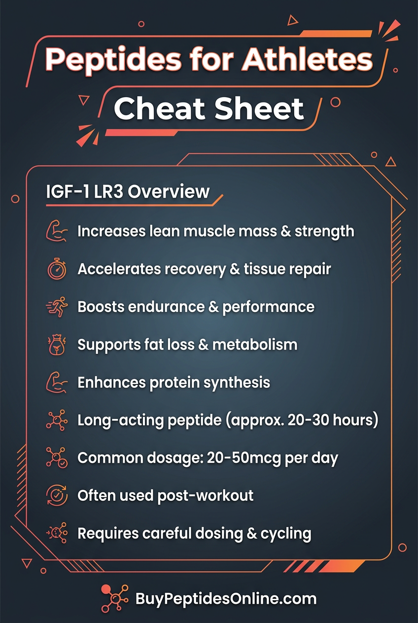
IGF-1 LR3: Half-life of approximately 20-30 hours — free and biologically active
This 60-90x increase in functional half-life transforms IGF-1 from a rapidly cleared, pulsatile signal into a sustained, continuous anabolic stimulus. A single administration of IGF-1 LR3 maintains biologically relevant concentrations for approximately 24 hours, making once-daily dosing feasible and producing a fundamentally different pattern of receptor activation than native IGF-1.
Distribution and Tissue Penetration
Because IGF-1 LR3 circulates largely unbound, it has access to interstitial spaces and tissue compartments that are less accessible to the heavily protein-bound native IGF-1. This enhanced tissue penetration is one reason why intramuscular injection of IGF-1 LR3 is theorized to produce localized effects — the compound can diffuse into surrounding muscle tissue and activate local IGF-1R populations before being cleared by local blood flow.
The volume of distribution of IGF-1 LR3 is considerably larger than that of native IGF-1, reflecting this enhanced tissue penetration. Research suggests that muscle tissue, adipose tissue, and liver all represent significant distribution compartments.
Clearance Mechanisms
IGF-1 LR3 is cleared primarily through receptor-mediated endocytosis (binding to IGF-1R and being internalized along with the receptor) and proteolytic degradation. Unlike native IGF-1, it does not rely on IGFBP-mediated transport for half-life extension, which means its clearance is less dependent on the body's IGFBP status — a variable that can change substantially with nutritional state, GH secretion, and other factors.
Enhanced Potency: The 2-3x Advantage
The combination of reduced binding protein sequestration and extended half-life makes IGF-1 LR3 approximately 2-3x more potent than native IGF-1 in cell proliferation assays, and potentially more potent still in whole-organism contexts where the extended half-life allows for sustained receptor occupancy. Researchers can explore lab-certified IGF-1 LR3 from verified suppliers to source material for such investigations.
---
Mechanisms of Muscle Growth: Hyperplasia, Hypertrophy, and Beyond
This section represents the heart of why IGF-1 LR3 has attracted such intense research interest in muscle biology. Its mechanisms are multifaceted, operating at the level of individual muscle fibers, satellite cell populations, and systemic metabolic regulation.
Hypertrophy: Making Existing Fibers Bigger
Hypertrophy — the enlargement of existing muscle fibers — is the most common form of skeletal muscle adaptation to anabolic stimuli. IGF-1 LR3 drives hypertrophy through several converging mechanisms:
PI3K/Akt/mTOR Pathway Activation:
The primary anabolic signaling cascade downstream of IGF-1R activation involves phosphoinositide 3-kinase (PI3K), which phosphorylates phosphatidylinositol-4,5-bisphosphate (PIP2) to generate phosphatidylinositol-3,4,5-trisphosphate (PIP3). PIP3 serves as a membrane anchor for Akt (protein kinase B), which is then phosphorylated and activated by PDK1 and mTORC2.
Activated Akt phosphorylates multiple downstream targets, but the most critical for muscle hypertrophy is mTORC1 (mechanistic target of rapamycin complex 1). mTORC1 activation drives:
S6K1 phosphorylation: → enhanced ribosomal biogenesis and translation initiation
4E-BP1 phosphorylation: → release of eIF4E and enhanced cap-dependent translation
Net effect: Dramatically increased rates of muscle protein synthesis
MAPK/ERK Pathway:
IGF-1R activation also engages the Ras/Raf/MEK/ERK pathway, which contributes to cell proliferation signaling and coordinates with mTOR to regulate protein synthesis. This pathway is particularly important for the proliferative (hyperplastic) effects of IGF-1 LR3 discussed below.
Anti-Catabolic Effects via FOXO Suppression:
Akt phosphorylates and inactivates the FOXO family of transcription factors, which are master regulators of muscle atrophy genes (atrogenes) including MuRF-1 and MAFbx/atrogin-1. By suppressing FOXO activity, IGF-1 LR3 simultaneously promotes anabolism and inhibits catabolism — a dual action that creates a strongly positive protein balance in muscle tissue.
Hyperplasia: The Unique Frontier
Hyperplasia — the actual creation of new muscle fibers through satellite cell activation and differentiation — is where IGF-1 LR3 becomes truly unique among anabolic research compounds. While most anabolic agents (including androgens, growth hormone, and mechanical loading) primarily drive hypertrophy of existing fibers, IGF-1 LR3's sustained activation of IGF-1R signaling appears capable of driving genuine myofiber hyperplasia through satellite cell biology.
This distinction is critical from a research standpoint — most anabolic agents only promote hypertrophy. The potential for genuine hyperplasia makes IGF-1 LR3 particularly interesting for physique and performance research, as well as for understanding the fundamental limits of muscle mass.
Satellite Cell Biology:
Muscle satellite cells are a population of adult stem cells that reside between the basal lamina and sarcolemma of muscle fibers. Under normal conditions, they are quiescent — maintained in a non-dividing state by a complex interplay of inhibitory signals. Following muscle damage, mechanical stress, or appropriate hormonal stimulation, satellite cells activate, proliferate, and ultimately differentiate into myoblasts that can fuse with existing fibers (contributing to hypertrophy) or fuse with each other to form entirely new myofibers (hyperplasia).
IGF-1 LR3's Role in Satellite Cell Activation:
IGF-1 LR3 activates muscle satellite cells through the PI3K/Akt/mTOR pathway described above, but also through:
Upregulation of MyoD and Myf5: — master transcription factors that commit satellite cells to the myogenic lineage
Stimulation of myogenin expression: — driving terminal differentiation of myoblasts
Promotion of myoblast fusion: — IGF-1R signaling enhances the expression of fusion-mediating proteins including myomaker and myomerger
Suppression of myostatin signaling: — IGF-1 LR3 indirectly antagonizes myostatin (a potent inhibitor of muscle growth) through Smad pathway interactions
The extended half-life of IGF-1 LR3 is particularly important for satellite cell biology. Satellite cell activation, proliferation, and differentiation are sequential processes that unfold over 24-72 hours. The brief pulse of native IGF-1 is insufficient to sustain this process through completion. IGF-1 LR3's 20-30 hour half-life provides the sustained signaling environment necessary to drive satellite cells through the full activation-proliferation-differentiation cycle.
Evidence for Hyperplasia:
Research in animal models has demonstrated that sustained IGF-1 overexpression (either through transgenic approaches or sustained administration of long-acting analogs) can produce genuine myofiber hyperplasia — a measurable increase in the number of individual muscle fibers, not just their size. While direct evidence in humans remains limited (as hyperplasia is difficult to measure non-invasively), the mechanistic basis is well-established, and IGF-1 LR3's pharmacokinetic profile makes it the most plausible candidate among available IGF-1 analogs for driving this effect.
Local vs. Systemic Effects
One of the most debated aspects of IGF-1 LR3 research is the question of local versus systemic action. Native IGF-1 is produced both hepatically (in response to GH) and locally within muscle tissue (in response to mechanical loading) — a distinction captured by the "somatomedin hypothesis" versus the "autocrine/paracrine IGF-1" model.
IGF-1 LR3, when administered intramuscularly into specific muscle groups, is theorized to exert preferential local effects on the injected tissue. The rationale:
1. High local concentrations immediately post-injection activate local IGF-1R populations
2. The compound diffuses into surrounding tissue before systemic clearance
3. Local satellite cell populations are exposed to sustained IGF-1R stimulation
This theoretical basis for site-specific injection protocols has made IGF-1 LR3 particularly interesting to researchers studying regional muscle hypertrophy and the autocrine/paracrine model of IGF-1 action. However, it should be noted that systemic distribution occurs regardless of injection site, and the relative contribution of local versus systemic effects remains an active research question.
---
Comparison with Related Compounds
Understanding IGF-1 LR3 in context requires comparing it to related molecules that operate in overlapping pharmacological spaces.
IGF-1 LR3 vs. Native IGF-1
| Feature | Native IGF-1 | IGF-1 LR3 |
|---|---|---|
| Half-life | 10-20 minutes | 20-30 hours |
| IGFBP binding | >95% bound | Minimal binding |
| Free fraction | <5% | ~95%+ |
| Receptor affinity | High | Slightly reduced but compensated |
| Net bioavailability | Low | Very high |
| Hyperplasia potential | Limited | Strong |
| Research convenience | Difficult (frequent dosing) | Practical (once daily) |
| Hypoglycemia risk | Moderate | High |
IGF-1 LR3 vs. IGF-1 DES
IGF-1 DES (des(1-3)IGF-1) is another modified IGF-1 analog, but it takes a different approach: rather than adding amino acids, it removes the first three amino acids from the N-terminus of native IGF-1. This truncation also reduces IGFBP binding, but to a lesser degree than LR3. IGF-1 DES has a shorter half-life than LR3 (approximately 20-30 minutes) but demonstrates dramatically enhanced receptor binding affinity — approximately 10x greater than native IGF-1 for the IGF-1R.
The practical consequence is that IGF-1 DES is better suited for acute, localized effects (particularly post-workout, when administered directly into target muscle), while IGF-1 LR3 provides sustained systemic anabolic stimulation. For a detailed comparison, see the dedicated IGF-1 DES vs IGF-1 LR3 comparison guide.
IGF-1 LR3 vs. MGF and PEG-MGF
Mechano Growth Factor (MGF) is a splice variant of the IGF-1 gene that is produced locally in muscle in response to mechanical damage. It operates primarily as a paracrine signal to activate satellite cells in the immediate vicinity of damage. PEG-MGF is a pegylated version with extended half-life.
MGF and IGF-1 LR3 have complementary but distinct mechanisms — MGF is primarily a satellite cell activator, while IGF-1 LR3 drives both satellite cell activation and the full downstream anabolic cascade. Research protocols sometimes combine these compounds for theoretically synergistic effects on muscle remodeling.
IGF-1 LR3 vs. GH Secretagogues
Growth hormone secretagogues — including GHRP-6, GHRP-2, ipamorelin, CJC-1295, and sermorelin — stimulate the pituitary to release endogenous GH, which then stimulates hepatic IGF-1 production. This is a fundamentally different approach: rather than providing exogenous IGF-1 directly, these compounds rely on the body's own GH/IGF-1 axis.
The advantages of the secretagogue approach include more physiological GH pulsatility, lower direct hypoglycemia risk, and a broader range of GH effects beyond IGF-1 (including direct GH receptor activation in adipose tissue). The disadvantage is that the IGF-1 response is indirect, variable, and subject to the constraints of endogenous IGFBP regulation.
IGF-1 LR3 bypasses all of this, delivering direct, sustained IGF-1R activation that is independent of GH secretion and IGFBP status.
Comprehensive Comparison Table
| Feature | IGF-1 LR3 | GH Peptides (GHRP+CJC) | Sermorelin | HGH | IGF-1 DES |
|---|---|---|---|---|---|
| Mechanism | Direct IGF-1R activation | Stimulate endogenous GH | Stimulate endogenous GH | Exogenous GH | Direct IGF-1R (local) |
| Hyperplasia | Yes (strong) | Weak/indirect | Weak/indirect | Weak/indirect | Yes (localized) |
| Half-life | 20-30 hours | Minutes to days | 10-20 minutes | 3-5 hours | 20-30 minutes |
| Hypoglycemia risk | High | Low | Low | Moderate | Moderate |
| IGFBP dependence | Minimal | High (endogenous IGF-1) | High (endogenous IGF-1) | High (endogenous IGF-1) | Low |
| Localized effects | Moderate | No | No | No | Strong |
| Cost | Moderate | Low | Low | High | Moderate |
| Research complexity | Moderate | Low | Low | Low | Moderate |
---
Dosing and Research Protocol Considerations
The following protocol information is provided for educational and research purposes only. These are not therapeutic recommendations.
Standard Research Dosing Parameters
Dose range:
Starting dose: 20 mcg per day (assess response, tolerance, and hypoglycemia risk)
Moderate research dose: 40 mcg per day
Higher research dose: 60-100 mcg per day (increased risk profile; used in some cell biology protocols)
Note: Doses above 100 mcg per day are associated with substantially increased side effect risk and are not commonly used in research protocols
Timing:
Post-workout administration: is the most common protocol rationale — the post-exercise window is characterized by elevated satellite cell responsiveness and enhanced IGF-1R expression in muscle tissue
Morning administration: is used in some protocols, particularly when post-workout timing is impractical
Divided dosing: (e.g., 20 mcg morning + 20 mcg post-workout) is used in some protocols to maintain more consistent receptor occupancy, though the 20-30 hour half-life makes this less pharmacokinetically necessary than with shorter-acting peptides
Route of administration:
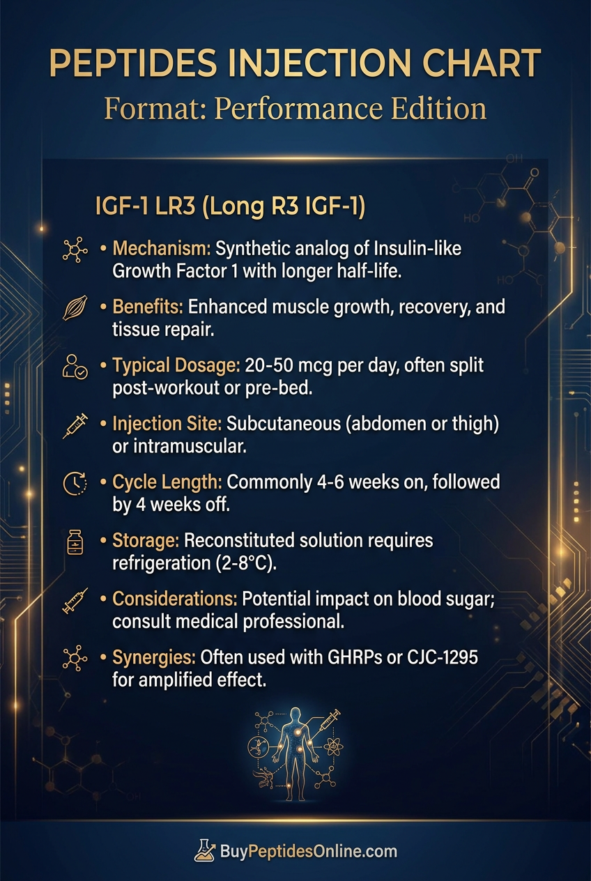
Subcutaneous injection: Standard approach; reliable absorption, predictable pharmacokinetics
Intramuscular injection into target muscle groups: Used in protocols specifically investigating localized hypertrophy or hyperplasia; bilateral injections into both sides of a target muscle group are common in such protocols
Intravenous administration: Used in some cell biology research contexts; not appropriate for standard research protocols
Cycle length:
Typical on-cycle: 4-6 weeks
Off-cycle: 4-6 weeks minimum
Rationale for cycling: IGF-1R downregulation (desensitization) occurs with sustained stimulation; cycling preserves receptor sensitivity and manages cumulative risk
Reconstitution and Handling
IGF-1 LR3 is typically supplied as a lyophilized (freeze-dried) powder, which must be reconstituted before use. Proper handling is essential for maintaining peptide integrity and research reproducibility.
Reconstitution protocol:
1. Allow the vial to reach room temperature before opening
2. Use bacteriostatic water (0.9% benzyl alcohol in sterile water) for reconstitution; this extends the shelf life of the reconstituted solution significantly compared to sterile water alone
3. Add the reconstitution solvent slowly to the side of the vial — do not inject directly onto the lyophilized powder
4. Gently swirl (do not shake vigorously) until the powder is fully dissolved; shaking can cause peptide aggregation and degradation
5. Standard reconstitution concentration: 1 mg in 2 mL bacteriostatic water = 500 mcg/mL; adjust based on desired injection volume
Concentration calculation example:
If reconstituting 1 mg (1000 mcg) in 2 mL bacteriostatic water:
Concentration = 500 mcg/mL
For a 40 mcg dose: draw 0.08 mL (80 µL) on an insulin syringe
Storage:
Lyophilized (unreconstituted): Store at -20°C for long-term storage; stable at 4°C for several months
Reconstituted solution: Refrigerate at 2-8°C; use within 4-6 weeks (bacteriostatic water extends this window vs. plain sterile water)
Avoid freeze-thaw cycling: of reconstituted solution — this degrades the peptide
Protect from light: IGF-1 LR3 is light-sensitive; store in amber vials or wrapped in foil
Never use if cloudy or particulate: — discard and use a fresh preparation
Injection Technique for Localized Research
For researchers investigating localized muscle effects:
Use a 28-31 gauge insulin syringe for intramuscular administration
Common target muscles for bilateral injection: biceps, triceps, quadriceps, deltoids
Inject slowly; aspirate before injection to confirm non-vascular placement
Rotate injection sites to prevent local tissue irritation
Some protocols specify injection immediately post-exercise into the worked muscle group
Blood Glucose Management
The hypoglycemic potential of IGF-1 LR3 is the most immediately relevant safety consideration in any research protocol. Practical blood glucose management:
Pre-injection: Ensure adequate carbohydrate availability; do not administer in a fasted state without monitoring
Post-injection monitoring: Blood glucose can decline significantly within 30-60 minutes of administration
Have fast-acting carbohydrates immediately available: Glucose tablets, fruit juice, or dextrose solution
Target blood glucose: Maintain above 70 mg/dL; below this threshold, hypoglycemia symptoms may emerge
Symptoms to monitor: Dizziness, sweating, tremor, confusion, rapid heartbeat
Timing with meals: Administering IGF-1 LR3 with or shortly after a carbohydrate-containing meal substantially reduces hypoglycemia risk
---
Stacking Considerations
IGF-1 LR3 is frequently studied in combination with other research compounds. The following combinations represent common research protocol designs.
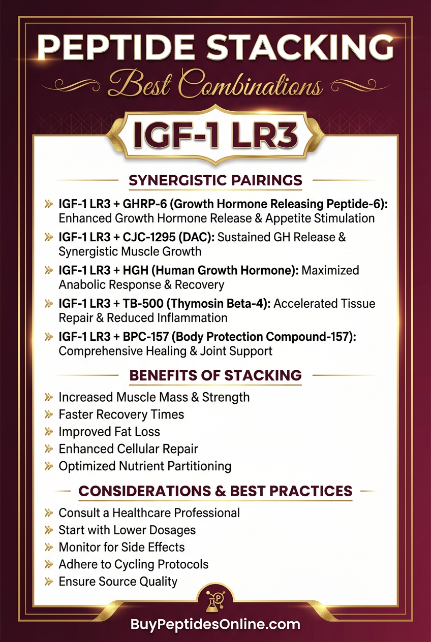
IGF-1 LR3 + GH Secretagogues
Combining IGF-1 LR3 with GH secretagogues such as ipamorelin + CJC-1295 creates a theoretically synergistic anabolic environment:
The secretagogue combination stimulates endogenous GH pulsatility, which drives hepatic IGF-1 production and activates GH receptors directly in muscle and adipose tissue
The combination addresses both GH receptor and IGF-1R signaling simultaneously
Research consideration: This combination significantly increases the complexity of the hormonal environment and the risk profile, particularly for hypoglycemia and IGF-1R-mediated proliferative effects.
IGF-1 LR3 + PEG-MGF
This combination targets complementary aspects of satellite cell biology:
PEG-MGF provides the initial satellite cell activation signal, mimicking the autocrine/paracrine MGF response to muscle damage
IGF-1 LR3 sustains the downstream anabolic cascade, driving the activated satellite cells through proliferation and differentiation
Some researchers sequence these compounds — PEG-MGF immediately post-workout followed by IGF-1 LR3 the following day — to mimic the natural temporal sequence of MGF and systemic IGF-1 responses to exercise.
IGF-1 LR3 + BPC-157
BPC-157 is a gastric pentadecapeptide with well-documented tissue repair, anti-inflammatory, and angiogenic properties. In research contexts, combining BPC-157 with IGF-1 LR3 is theorized to:
Accelerate repair of connective tissue and vasculature to support the hypertrophic response driven by IGF-1 LR3
Reduce exercise-induced inflammation that might otherwise limit training stimulus
Enhance the local angiogenic environment necessary to support new muscle fiber growth
IGF-1 LR3 + TB-500
TB-500 (Thymosin Beta-4) promotes actin polymerization, wound healing, and tissue regeneration. Like BPC-157, it is studied as a complementary recovery-supporting compound in protocols where IGF-1 LR3 is driving aggressive muscle remodeling.
IGF-1 LR3 + Follistatin-344
Follistatin-344 is a myostatin antagonist — it binds and neutralizes myostatin, the primary endogenous inhibitor of muscle growth. The theoretical rationale for combining with IGF-1 LR3 is powerful:
IGF-1 LR3 maximally activates the anabolic signaling cascade (PI3K/Akt/mTOR, satellite cell activation)
Follistatin-344 removes the primary brake on muscle growth (myostatin inhibition)
The combination theoretically creates a maximally permissive environment for muscle hyperplasia and hypertrophy
This combination is among the most aggressive in the research peptide space and carries a correspondingly elevated risk profile.
---
Safety Profile and Documented Side Effects
IGF-1 LR3 has a well-characterized side effect profile based on decades of research with IGF-1 analogs. Understanding these risks is essential for responsible research protocol design.
Hypoglycemia: The Primary Acute Risk
As noted above, hypoglycemia is the most immediately dangerous acute risk associated with IGF-1 LR3. The mechanism involves:
Direct activation of IR-A (insulin receptor isoform A) by IGF-1 LR3, which shares structural homology with insulin
IGF-1R-mediated glucose uptake in muscle and adipose tissue
Potential suppression of hepatic glucose output
The hypoglycemic effect of IGF-1 LR3 is dose-dependent and is substantially more pronounced than with native IGF-1 due to the extended half-life — a hypoglycemic episode can be prolonged rather than brief. Researchers must have glucose management protocols in place before beginning any IGF-1 LR3 research.
Receptor Downregulation and Desensitization
Sustained IGF-1R activation leads to receptor internalization and downregulation — a process called homologous desensitization. With the 20-30 hour half-life of IGF-1 LR3, this process can be significant over multi-week research periods. This is the primary pharmacological rationale for cycling protocols (4-6 weeks on, 4-6 weeks off), which allow receptor populations to recover and restore sensitivity.
Visceral and Organ Growth Concerns
IGF-1R is expressed in virtually all tissues, including the gastrointestinal tract, heart, kidneys, and other visceral organs. Sustained IGF-1R activation can theoretically drive growth in these tissues as well as in skeletal muscle. The most commonly discussed concern is gut growth — thickening of the intestinal wall — which has been observed in animal models with chronic IGF-1 overexpression. This concern is primarily theoretical at research doses and cycle lengths, but represents a genuine long-term risk with chronic, high-dose use.
Acromegaly-Like Effects
Acromegaly — the clinical syndrome caused by chronic GH/IGF-1 excess — is characterized by enlargement of the jaw, hands, feet, and facial features, as well as internal organ enlargement and metabolic dysfunction. These effects require sustained, supraphysiological IGF-1 exposure over months to years to develop. At research doses and cycle lengths, this risk is theoretical rather than practically demonstrated, but it represents a genuine concern with chronic abuse.
Tumor Promotion
This is perhaps the most serious long-term safety concern with IGF-1 LR3. IGF-1R signaling is a potent driver of cell proliferation and survival — it is upregulated in numerous cancer types and has been identified as a therapeutic target in oncology. Sustained IGF-1R activation by IGF-1 LR3 could theoretically:
Accelerate the growth of pre-existing tumors
Promote the survival of pre-malignant cells
Enhance angiogenesis that supports tumor growth
IGF-1 LR3 is absolutely contraindicated in research subjects with active or recent cancer history. This is a non-negotiable safety boundary.
Edema and Water Retention
Like native IGF-1 and GH, IGF-1 LR3 can cause fluid retention, particularly in the early weeks of use. This manifests as peripheral edema (swelling in the extremities) and joint discomfort. This effect is generally dose-dependent and resolves with dose reduction or discontinuation.
Fatigue and Lethargy
Some research subjects report fatigue, particularly in the first 1-2 weeks of IGF-1 LR3 use. The mechanism is not fully elucidated but may relate to metabolic shifts, mild hypoglycemia, or the significant anabolic demands placed on the body during active IGF-1R stimulation.
Injection Site Reactions
Local injection site reactions — redness, mild swelling, discomfort — are common with any peptide injection protocol and are generally mild and transient. Proper injection technique, site rotation, and attention to sterility minimize these effects.
---
Common Research Mistakes
Based on the accumulated research experience with IGF-1 LR3, several protocol errors are particularly common and consequential:
1. Failing to manage blood glucose:
The single most dangerous mistake. Administering IGF-1 LR3 in a fasted state without glucose monitoring or readily available carbohydrates has led to significant hypoglycemic events in research contexts.
2. Starting at too high a dose:
Beginning at 60-100 mcg rather than the recommended starting dose of 20 mcg prevents assessment of individual sensitivity and dramatically increases side effect risk.
3. Skipping cycle breaks:
Using IGF-1 LR3 continuously without off-cycle periods leads to receptor desensitization, diminishing returns, and cumulative risk accumulation.
4. Improper reconstitution:
Injecting bacteriostatic water directly onto the lyophilized powder (rather than the vial wall), shaking vigorously, or using plain sterile water (which shortens shelf life) can degrade the peptide and compromise research results.
5. Inadequate cold chain maintenance:
Allowing reconstituted IGF-1 LR3 to reach room temperature repeatedly or exposing it to light degrades the peptide and reduces potency.
6. Ignoring IGFBP context:
Native IGF-1 levels and IGFBP status vary substantially with nutritional state, GH secretion, and health status. While IGF-1 LR3 is less dependent on IGFBP context than native IGF-1, researchers should be aware that the overall IGF-1 signaling environment is not determined solely by exogenous LR3 administration.
7. Sourcing from unverified suppliers:
IGF-1 LR3 purity and accurate concentration are critical for reproducible research. Third-party tested material from verified suppliers is essential. Explore lab-certified IGF-1 LR3 from verified suppliers to ensure research-grade quality.
8. Using in contraindicated subjects:
Administering IGF-1 LR3 in research contexts involving subjects with active cancer, severe diabetes, or other conditions where IGF-1R stimulation is contraindicated represents a serious ethical and safety breach.
---
Who Is IGF-1 LR3 Studied In?
From a research perspective, IGF-1 LR3 has been employed across a remarkably diverse range of investigational contexts:
Muscle biology research:
The primary research application. IGF-1 LR3 is used to study satellite cell biology, myofiber hyperplasia, protein synthesis regulation, and the molecular mechanisms of muscle hypertrophy. Its extended half-life makes it far more practical than native IGF-1 for sustained in vivo experiments.
Cancer biology:
IGF-1R signaling is extensively studied in oncology, and IGF-1 LR3 is used as a research tool to activate IGF-1R in cancer cell lines and animal models, studying proliferation, survival, migration, and response to IGF-1R-targeted therapies.
Metabolic research:
IGF-1's role in insulin sensitivity, glucose metabolism, and adipose tissue regulation makes IGF-1 LR3 useful in metabolic disease research, including studies of insulin resistance and GH deficiency states.
Aging research:
IGF-1 levels decline with age (a phenomenon called "somatopause"), and IGF-1 LR3 is studied in the context of age-related muscle loss (sarcopenia), bone density decline, and metabolic deterioration.
Wound healing and tissue repair:
IGF-1R signaling promotes the proliferation and migration of multiple cell types involved in wound healing, including fibroblasts, keratinocytes, and endothelial cells. IGF-1 LR3 is studied in this context as a sustained stimulus for tissue repair.
Performance and physique science:
In the research peptide space, IGF-1 LR3 is primarily studied for its effects on skeletal muscle mass, body composition, and recovery from exercise-induced muscle damage — the focus of this article.
---
Research Outlook: Where Is IGF-1 LR3 Science Heading?
The research landscape for IGF-1 LR3 and related compounds is evolving rapidly, driven by advances in our understanding of IGF-1R biology, satellite cell regulation, and the molecular mechanisms of muscle growth.
Mechanistic Refinement
Ongoing research is clarifying the precise contributions of IGF-1 LR3 to each phase of satellite cell biology — activation, proliferation, differentiation, and fusion. Single-cell RNA sequencing and advanced imaging techniques are allowing researchers to track individual satellite cells through these processes in response to IGF-1R stimulation, providing unprecedented mechanistic resolution.
Hyperplasia in Humans: The Open Question
Perhaps the most important unresolved question in IGF-1 LR3 research is the extent to which genuine myofiber hyperplasia occurs in adult humans. Animal models clearly demonstrate hyperplasia with sustained IGF-1 overexpression, but human muscle biopsy studies have produced mixed results. Advances in muscle fiber counting techniques (including non-invasive MRI-based approaches) may eventually resolve this question.
Combination Therapy Research
The potential for IGF-1 LR3 to complement other anabolic and regenerative compounds — particularly myostatin inhibitors like follistatin and anti-myostatin antibodies — is an active research frontier. The theoretical synergy between removing the brake on muscle growth (myostatin inhibition) and maximally activating the anabolic accelerator (IGF-1R stimulation) represents one of the most compelling areas of muscle biology research.
Sarcopenia and Aging Applications
As the global population ages, sarcopenia — the progressive loss of muscle mass and function with aging — is recognized as a major public health challenge. IGF-1 LR3, with its ability to activate satellite cells and drive muscle protein synthesis even in the face of age-related anabolic resistance, is studied as a potential tool in this context. Clinical translation faces significant regulatory and safety hurdles (particularly the tumor promotion concern), but preclinical results are promising.
Improved Analogs
Research into next-generation IGF-1 analogs — with even more refined receptor selectivity, tissue targeting, or modified clearance profiles — is ongoing. Some researchers are exploring biased agonism at the IGF-1R (selectively activating certain downstream pathways while avoiding others) as a strategy to preserve anabolic effects while reducing proliferative risks.
For a broader perspective on muscle-building peptides in the current research landscape, the best peptides for muscle growth 2026 guide provides comprehensive context.
---
Frequently Asked Questions
What is the difference between IGF-1 LR3 and regular IGF-1?
Native IGF-1 is a 70-amino acid peptide produced primarily in the liver that is rapidly cleared from circulation (half-life 10-20 minutes) and extensively bound by IGF Binding Proteins (over 95% bound at any time). IGF-1 LR3 is an engineered analog with two structural modifications — a 13 amino acid N-terminal extension and an arginine substitution at position 3 — that together dramatically reduce IGFBP binding affinity while preserving IGF-1 receptor affinity. The result is a molecule with a half-life of 20-30 hours that circulates largely free and biologically active, making it approximately 2-3x more potent than native IGF-1 in cell proliferation assays and far more practical for research protocols requiring sustained IGF-1R activation.
Why is the 20-30 hour half-life of IGF-1 LR3 so significant?
The extended half-life transforms IGF-1 LR3 from a brief, pulsatile signal into a sustained anabolic stimulus. This matters enormously for satellite cell biology — the sequential processes of satellite cell activation, proliferation, and differentiation unfold over 24-72 hours, and a brief pulse of native IGF-1 is insufficient to sustain this process through completion. IGF-1 LR3's extended half-life provides the continuous receptor occupancy necessary to drive satellite cells through the full cycle, which is the mechanistic basis for its potential to drive genuine myofiber hyperplasia. For researchers, the extended half-life also means once-daily dosing is pharmacokinetically appropriate, simplifying protocol design.
Can IGF-1 LR3 actually create new muscle fibers (hyperplasia)?
This is one of the most important and contested questions in IGF-1 LR3 research. In animal models — including rodents and various in vitro systems — sustained IGF-1R activation clearly promotes satellite cell-mediated myofiber formation, and genuine increases in myofiber number have been demonstrated with chronic IGF-1 overexpression. The mechanistic basis is well-established: IGF-1R activation drives satellite cell activation, proliferation, and differentiation into new myofibers. Whether this translates to meaningful hyperplasia in adult humans at research doses remains an open question, primarily because of the difficulty of measuring myofiber number non-invasively in humans. The mechanistic plausibility is strong, and IGF-1 LR3 remains the most pharmacokinetically appropriate tool for investigating this question.
What is the hypoglycemia risk with IGF-1 LR3, and how should it be managed?
Hypoglycemia is the most immediately significant acute risk with IGF-1 LR3. The compound activates both IGF-1 receptors and, to a lesser degree, insulin receptors (particularly the IR-A isoform), driving glucose uptake in muscle and adipose tissue while potentially suppressing hepatic glucose output. The extended half-life means that hypoglycemia can be prolonged rather than brief. Management strategies include: never administering in a fasted state, consuming carbohydrates around injection time, having fast-acting glucose sources immediately available, monitoring blood glucose post-injection (particularly in the first 30-90 minutes), and starting at the lowest research dose (20 mcg) to assess individual sensitivity before escalating.
How should IGF-1 LR3 be cycled, and why?
Research protocols typically use IGF-1 LR3 for 4-6 weeks followed by 4-6 weeks off. The primary rationale is receptor biology: sustained IGF-1R activation leads to receptor internalization and downregulation (homologous desensitization), which reduces the anabolic response over time. Cycling allows IGF-1R populations to recover and restore sensitivity, preserving the efficacy of subsequent research cycles. Cycling also manages cumulative risk — limiting total exposure to IGF-1 LR3 reduces the theoretical risks associated with sustained IGF-1R activation, including proliferative effects in non-target tissues.
What compounds are commonly studied alongside IGF-1 LR3?
IGF-1 LR3 is studied in combination with a range of complementary compounds. GH secretagogues (ipamorelin, CJC-1295) are combined to stimulate endogenous GH pulsatility alongside direct IGF-1R activation. PEG-MGF is combined to target the initial satellite cell activation phase that IGF-1 LR3 then sustains. BPC-157 and TB-500 are added for connective tissue repair support. Follistatin-344 is combined to remove myostatin-mediated inhibition of muscle growth. Each combination adds complexity and risk, and should be approached with appropriate caution in research contexts.
Where can I source research-grade IGF-1 LR3?
Research-grade IGF-1 LR3 should be sourced from suppliers who provide third-party analytical testing (HPLC purity, mass spectrometry confirmation of molecular identity, and sterility testing). Accurate concentration and high purity are essential for reproducible research results and for accurate dose-response characterization. For guidance on identifying legitimate research-grade suppliers, the dedicated where to buy IGF-1 LR3 online guide provides detailed sourcing criteria and vendor evaluation frameworks. Third-party tested IGF-1 LR3 is available from trusted research vendors who meet these quality standards.
---
Key Takeaways
IGF-1 LR3 occupies a unique and compelling position in the landscape of research peptides. Its structural modifications — a seemingly simple N-terminal extension and single amino acid substitution — produce pharmacokinetic consequences that are anything but simple, transforming a rapidly cleared, extensively bound native hormone into a sustained, highly bioavailable anabolic signal.
The core takeaways for researchers:
IGF-1 LR3 is the most potent form of IGF-1 available for research: , combining reduced IGFBP binding with a 20-30 hour half-life to deliver sustained IGF-1R activation that native IGF-1 cannot achieve
Its extended half-life is not merely a convenience: — it is mechanistically essential for driving satellite cell biology through the full activation-proliferation-differentiation cycle that underlies potential myofiber hyperplasia
Unique potential for muscle hyperplasia: sets it apart from virtually all other anabolic research compounds, which primarily drive hypertrophy of existing fibers
The PI3K/Akt/mTOR pathway: is the central anabolic signaling cascade activated by IGF-1 LR3, with additional contributions from MAPK/ERK signaling and FOXO suppression creating a strongly anabolic, anti-catabolic cellular environment
Careful blood glucose management is non-negotiable: — hypoglycemia is the most immediately dangerous acute risk and must be actively managed in any research protocol
Cycling (4-6 weeks on, 4-6 weeks off) is pharmacologically rational: , preserving IGF-1R sensitivity and managing cumulative risk
Sourcing quality matters enormously: — research reproducibility and safety both depend on accurate concentration and high purity; third-party tested IGF-1 LR3 is available from trusted research vendors
The tumor promotion concern is real and serious: — IGF-1 LR3 is absolutely contraindicated in research contexts involving subjects with active or recent cancer
The research frontier for IGF-1 LR3 remains active and exciting. Questions about the extent of genuine hyperplasia in adult humans, the optimal combination protocols for maximizing muscle remodeling, and the long-term safety profile of sustained IGF-1R activation are all subjects of ongoing investigation. As our mechanistic understanding deepens — particularly with advances in single-cell biology and non-invasive muscle imaging — the picture of what IGF-1 LR3 can and cannot do will become progressively clearer.
For now, it remains one of the most scientifically interesting and mechanistically distinctive research peptides available — a molecule that asks fundamental questions about the limits of muscle biology and provides a uniquely powerful tool for investigating the answers.
---
Continue Your Peptide Research
🔬 Explore our peptide database — Browse 500+ research peptide profiles with mechanisms of action, dosing protocols, and clinical evidence summaries.
🤖 Have questions? — Ask PeptideAI, our research assistant, for personalized peptide guidance based on the latest studies.
📚 Want more guides? — Browse all research articles covering peptide science, comparisons, and buying guides.
Related Articles on BuyPeptidesOnline.com
Continue your research with these in-depth guides: